Every person has uniquely-shaped ears that continue to grow over time. Making an accurate reproduction of the ear is an important part of delivering a customised ear product. From the early 1950s, this was achieved using a paste which set in the ear, a technique which still underpins common practice today. However, advancements in laser technology and 3D virtual images are shaping the future of ear products. These developments have a variety of potential applications that may offer improvements in many areas including custom devices for hearing, music, communications, protection and the field of biometrics.
In this spotlight we talk to Dana Helmink, the global product manager of OTOSCAN, who introduces us to an innovative ear scanning solution. We also hear a patient’s experience of having a silicone ear impression taken and the student audiologist’s experience of assessing a variety of traditional silicone based techniques for the first time.
A patient’s story:
Hello Caspian. You have had casts of both ears taken today with silicone impression material. I wonder if you could share your experience with us?
Firstly, cotton buds on strings were lodged into my ears as far as possible. This can feel a bit uncomfortable because it feels like they are nearly touching the ear drum. I knew it was being done by an audiologist so I had no fear of damage to the ear drum. Next phase was to fill the ears with a syringe of thick paste. It was a bit of a strange sensation but not at all uncomfortable. Then everything goes quite, which was quite nice in this hectic noisy world we live in. It took about five minutes for the impressions to set, they were removed and it was all over. Rather quick and non-eventful.
A student audiologist story:
Hello Christine. You’ve just completed a clinical tutorial on silicone impression taking and completed your first ear impression on a patient. Could you share your experiences with us?
During the tutorial I practised using three different types of delivery system including a few styles of manual syringes, a manual cartridge gun and an electronic cartridge gun. I found experimenting with the various types of equipment beneficial to help explore the advantages and disadvantages of each. During the tutorial I felt the manual syringe was most easily controlled because it was the easiest to brace with. The challenging aspect was limiting small air pockets and folds in the impression material as it flowed. This was perhaps due to initial inconsistent pressure and movement while taking the impression. I improved on this technique with practice.
The manual cartridge gun I disliked because it was difficult to repeatedly squeeze and my hand felt a bit sore after use. The inconsistent flow of material left the impression of poor quality during my first attempts. The cartridge gun also made a clicking noise when the material was squeezed. The gun was larger than the manual syringe. Both these points did not appeal to me as I felt patients might not be at ease with the noise and squeezing motion. This in turn might have caused possible jaw tension and movement. I did not have confidence in this technique.
The electronic gun resulted in a high quality impression, however I found it difficult to brace because it was much larger than the syringe. The machine delivered the flow of the silicone quicker into the ear. I felt the size of the equipment might alarm some patients and possibly cause harm if the clinician was unable to brace effectively. The mixing of the material was great because it mixed accurate quantities and was very hygienic since the material was not as exposed like the manual syringe.
After my tutorial and extra reading of the recommended procedures I took my first impression on a patient. This impression was for a custom ear piece for a hearing aid. The patient was a dentist and he was very friendly and happy for me to go ahead under supervision. When I was doing the impression I was terrified that I was going to hurt the patient or cause discomfort. I prepped the patient and got the otostop and impression material ready before I started. When I placed the otostop into the ear I noticed that there was a small gap at the bottom of the ear canal. I repositioned it so it was safe to proceed. I used the manual syringe by choice. I found it challenging to confirm that I was applying the correct amount of pressure consistently to eliminate air bubbles and folds in the material, however I managed the procedure safely and braced easily. After I had finished and the impression was removed the patient said immediately “I have my hearing back and can talk again”. This made me realise how isolating the experience of getting an impression can be.
There are many well-known factors that contribute to variability in the process of taking an ear impression and delivering a great fitting ear piece. Variables include the syringe system, style and placement of otoblock, material viscosity, shore value, clinical technique, shrinkage, packaging and the manufacturing process. In principle a 3D scan allows a true reproduction of the ear and does not contain the same inherent variability of traditional methods. Next, we hear from Dana about the new OTOSCAN development.
Q&A with Dana Helmink, Au.D. Global Product Manager, OTOSCAN; GN Otometrics
Can you tell us a little about how the OTOSCAN system works?
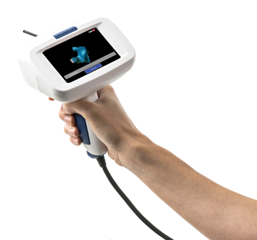
The OTOSCAN is a complete earscanning solution consisting of a handheld scanner, laptop computer, patient worn tracking headset, and web-based ordering portal designed to replace the current silicone impression process.
The ear is a complex shape with sharply curved surfaces, some essentially flat surfaces, and narrow openings. While 3D scanning has been used in many applications for many years, the ear has remained uncharted territory due to these challenges. The OTOSCAN is uniquely designed to address these challenges with a solution that supports scanning all surfaces of the ear from the full pinna past the second bend of the ear canal.
When scanning the ear, laser light is emitted by either the ring laser located in the probe tip or the fan laser located at the base of the probe. The laser is visualised on the surface of the skin by a miniature camera also contained within the probe tip. As the camera captures the image of the laser on the surface of the skin, sophisticated algorithms convert the image into data points and the tracking system, consisting of the tracker cameras and patient-worn headset, stitches the data points together into a 3D image of the ear. This patented system breaks the barrier for earscanning.
Can you describe the procedure from a patient perspective?
The patient experience is best described as comfortable and interactive. The OTOSCAN makes little or no contact with the ear so the discomfort related to placing an otoblock is eliminated as is the pressure and occlusion related to injecting silicone material. During the scan the patient will be seated in a chair that provides easy access to the ears and will be able to view the scan process and 3D image on the computer display. Scanning can be paused and resumed as necessary to allow for interaction between the patient and clinician. It’s a world of difference from the current silicone impression process.
Is it quicker and safer than traditional methods of taking ear impressions?
Yes, two of the most notable benefits of the OTOSCAN are that it offers a safer and more efficient process compared to the traditional silicone impression process. A complete scan of the outer ear and canal can be completed in 2-3 minutes. At the end of scanning, the 3D image is automatically uploaded to the web-based portal where the clinician can complete the order and instantly submit to the manufacturer for processing. Shipping and handling times are reduced throughout the process.
To improve patient safety, the scanner’s tracking system provides a measure of the insertion depth into the ear canal and the built-in display includes a live view of the ear. Together these features support safe navigation in the ear canal. The risk of silicone bypassing the otoblock is eliminated and reaching the desired canal length is no longer an act of trial and error.
How does the system manage artefacts such as cerumen and hair?
Since hair, cerumen and skin all have different optical traits, we’ve been able to develop specialised optical systems and advanced software algorithms that are able to ignore hair and reasonable amounts of cerumen.
Is it safe to scan surgically modified ears such as mastoid cavities?
Again, you have hit on one of the great advantages offered by earscanning with OTOSCAN. Surgically altered ears present a significant challenge and risk with the traditional silicone impression process. The OTOSCAN makes minimal contact with the ear during the scanning process and the clinician can navigate the ear canal safely using the video otoscope and there is, of course, no risk that impression material will become stuck in the mastoid cavity.
Ear canals change shape when chewing, yawning and smiling etc. How does the system take into account the elasticity of the ear canal?
The OTOSCAN makes it easy to take both a closed jaw and open jaw scan without the need to wait through the cure time involved with two sets of silicone impressions or perform duplicate tedious steps, such as placing an otoblock. Additionally there is no added materials cost in taking two scans of the ear. It’s fair to say that OTOSCAN makes it practical to scan the ear in both conditions.
What age is the procedure suitable for?
At its release the OTOSCAN will be intended for use on patients with adult-sized ear canals and heads which means that adults and older children can be scanned. Support for scanning younger paediatric patients will be part of future releases and development.
Is specialist equipment required?
The OTOSCAN system includes everything necessary for scanning and will be available under a subscription / lease agreement with simple pay-per-order payment plan. No direct investment in hardware and software will be required.
When will the system be available?
Please sign up at www.earscanning.com to be the first to hear the news regarding the launch of this remarkable new technology.
Thanks to Dana Helmink, Caspian Guirey and Christine McDermott for taking time to contribute to this spotlight.
This product offers a new solution that may transform the ear impression process. It unlocks future benefit for the ear industry including efficiency in practice, reduced quantity of remakes, true beyond the bend reproduction of the ear, enhanced safety and standards, reduction of impression material waste and postal burden. We look forward to hearing more about the upcoming launch and the future developments.
‘Spotlight on Innovation’ is an informative section to provide insight and discussion on recent advances in technology and research and does not imply endorsement by ENT and Audiology News.