Although China was late to establish head and neck surgery in comparison to other areas of oncological surgery, with the persistent hard work of head and neck surgeons nationally, it has progressed significantly during the last two decades. Yueying Ma and Liangfa Liu discuss this development.

Beijing Friendship Hospital, Capital Medical University.
Head and neck as a separate surgical specialty was established in China in the early 1950s. The professional commission of head and neck surgery was formally constituted in 1985. Most head and neck surgeons in our country work almost exclusively in dedicated departments of head and neck surgery at cancer hospitals or oral and maxillofacial surgery of general hospitals, or oral and maxillofacial surgery in hospitals of stomatology (or dental hospitals). Each of these departments is complementary and has its respective contributions.
In China, we have tried to keep up with the cutting edge techniques practised in the leading centres of the world, including multidisciplinary treatment of head and neck cancer, skull-base tumour surgery, laryngeal preservation in laryngeal carcinoma and hypopharyngeal carcinoma, functional reconstruction of head and neck after radical tumour resection, and minimally invasive head and neck surgery [1]. During the past 20 years, with the introduction of new technology and techniques, head and neck surgery in China has developed rapidly throughout the country.
Based on the death survey data of 2008 from the Cancer Hospital of Chinese Academy of Medical Sciences (age-standardised incidence rates) head and neck cancer was in the top 20 cancers estimated in China in 2008 including: esophageal cancer (fourth in male and seventh in female); nasopharyngeal cancer (11th in male); laryngeal cancer (14th in male); and thyroid cancer (13th in female) [2]. Based on the data of the latest cancer statistics in 2015 from the same hospital, 477,900 new esophageal cancers and 90,000 new thyroid cancers were estimated in 2015 [3]. This shows that currently laryngeal, thyroid and esophageal malignancies are still the main focus of head and neck surgery.
“Like in the west, transoral laser microsurgery for early laryngeal cancer has been adopted widely at different head and neck centres in China.”
Radical surgery for head and neck cancer has often resulted in organ dysfunction, morbidity and disfigurement but with increasing economic prosperity, patients now aspire to better quality of life. The head and neck surgeons have to pay more attention to preservation of function and reconstruction of organs, along with radical resection. Surgery with laryngeal preservation and functional reconstruction is being practised for advanced laryngeal and hypopharyngeal carcinomas. Orbital preservation and function has to be taken into account in paranasal sinus cancer operations. Reconstruction of the maxilla and mandible with flaps and dental implants are widespread in maxillofacial oncological surgery.
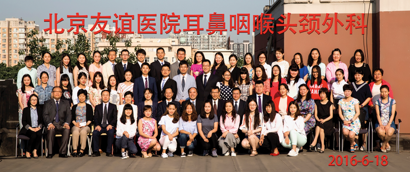
Staff of the department of Otolaryngology, Head Neck Surgery, Beijing Friendship Hospital, Capital Medical University.
Multi-centre studies have shown that open laryngeal surgery, laser microlaryngeal surgery and primary radiotherapy have similar survival rates for early glottic cancer in our country. Like in the west, transoral laser microsurgery for early laryngeal cancer has been adopted widely at different head and neck centres in China [4]. For T2 glottic laryngeal cancer, vertical partial laryngectomy could get about a 90% five-year postoperative survival rate and an 80-100% decannulation rate [5]. Supracricoid partial laryngectomy (with cricohyoidoepiglottopexy) could get an 88-90% five-year postoperative survival rate and a 98-100% decannulation rate [6]. Some laryngeal preservation surgery for advanced laryngeal cancers has also achieved good outcomes.
Radial forearm free flaps and free anterolateral thigh flaps have been used to repair supraglottic structures for laryngeal preservation after partial laryngectomy in patients with advanced hypopharyngeal carcinoma [7]. We have reported a number of cases in which repairs of non-circumferential defects were carried out with a submental island flap after hypopharyngeal carcinoma ablation, and got satisfactory results [8]. For early hypopharyngeal carcinoma, there are also some attempts of coblation applications [9].
In recent years, transoral robotic surgery is establishing itself internationally as a new tool in performing complex resections in head and neck surgery, and some hospitals in our country have also developed this new technology as an application in thyroid gland, oropharyngeal and hypopharyngeal surgery [10], and received satisfactory outcomes.
In rhinology, endoscopic sinus surgery has been applied for benign disease of the nasal cavity, paranasal sinuses and skull-base for nearly three decades in Europe and North America. In recent years, surgeons in our country have tried to use endoscopic sinus surgery in most paranasal benign tumours, including sinonasal inverted papillomas, nasopharyngeal angiofibroma, and even some localised intranasal and sinus carcinomas. Endoscopic skull base surgical resection and reconstruction has also developed over the last decade with excellent outcomes. In some large tertiary head and neck centres, image-guided systems are used in conjunction with endoscopic sinus surgery to recognise and preserve important structures, reduce complications, ensure good tumour excision margins, and improve total resection rate along with reduced postoperative morbidity [11].
Three-dimensional rapid prototyping is a new technique which has been used and developed in many medical fields and is especially popular in maxillofacial head and neck surgery, including operation planning, simulated operation and making individualised implant prosthesis. In China, Sun first attempted to reconstruct the maxilla and mandible with supplementary three-dimensional rapid prototyping, and has reported more than one hundred such procedures so far [12,13].
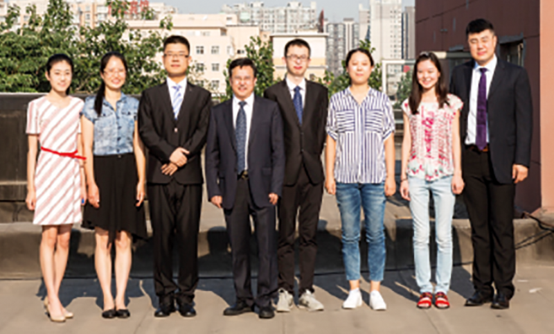
Staff of the Subdivision of Surgery, Beijing Friendship Hospital, Capital Medical University.

Multidisciplinary team (MDT) meeting.
Reducing disabling morbidity associated with cranial nerve paresis during head and neck tumour resection is a key objective of most head and neck surgeons. In recent years, intraoperative cranial nerve monitoring is widely being applied in head and neck surgery in an attempt to improve surgical outcomes. Wang reported his successful preservation of the facial nerve function during parotid gland surgery with intraoperative facial nerve monitoring [14]. Recurrent laryngeal nerve and vagus nerve monitoring is also being increasingly applied in thyroid gland surgery in many institutions, and helps with training and improved outcomes. In endoscopic optic nerve decompression, sinus endoscopic optic surgery involving orbit and anterior skull base, intraoperative optic nerve monitoring is helping to avoid optic nerve injury [15].
A new generation of lymphatic tracers, nanocrystalline carbon has been widely used in surgery of gastric cancer and breast cancers, guiding lymph node resection and sentinel lymph node biopsy. In recent years, nanocrystalline carbon tracers have been applied in thyroid gland surgery. Zhang found that there are different lymphatic drainage pathways between thyroid glands and parathyroid glands, and nanocrystalline carbon tracing could help improved localisation of parathyroid glands [16].
Zhu and his colleagues reported that they could help preserve parathyroid glands and the vagus nerve, and thus reduce complications and plan lymph node dissection more accurately and completely with nanocrystalline carbon tracer application in thyroid gland surgery [17]. Some surgeons also apply nanocrystalline carbon into sentinel lymph node mapping in head and neck cancer. Ren and his colleague used nanocrystalline carbon mixed suspension to detect sentinel lymph node in 20 cN0 laryngeal cancer operations, and in 19 out of 20 displayed sentinel lymph node successfully (detection rate is 95%) [18]. Guan et al injected nanocrystalline carbon mixed suspension in the margin of tumour preoperatively in 45 patients with oral squamous cell carcinomas, and guiding cervical lymph node dissection with lymph node black staining [19].
“In China, Sun first attempted to reconstruct the maxilla and mandible with supplementary three-dimensional rapid prototyping, and has reported more than one hundred such procedures so far.”
The history of radioactive seed implantation in treatment of cancer is more than 100 years in Europe and USA. Our scientists developed iodine seeds (model 6711) and applied them in clinical practice over the last two decades. The application and approach of radioactive seed implantation in head and neck surgery has also been applied selectively. In 2006, Zhang and colleagues treated 36 cases of oral and maxillofacial malignancy with CT-guided iodine, seed implantation and followed for 6 to 36 months, with no recurrence founded in target region [20]. In 2010, Qiao et al treated 45 cases of oral and maxillofacial malignant tumours (47 lesions) with iodine125 seed implantations under ultrasound-guidance, and the application was generally very accurate [21]. In 2014, Feng and colleagues first implanted the iodine125 seed with the guidance of a three-dimensional printing model for a patient with a maxillary sinus tumour. Compared with CT or ultrasound guidance, the three-dimensional printing model meant less operative time and improved accuracy with better effects [22].
The management of head and neck malignancy needs multidisciplinary cooperation, and improving survival rate and pursuing quality of life at the same time is our ultimate objective. At present in China, most of the head and neck centres have set up multidisciplinary consultation procedures, that utilise multiple current management modalities (including surgery, radiotherapy, chemotherapy, bio-immunotherapy, gene therapy and Chinese traditional treatment) comprehensively in a planned way, in order to improve cure rate and quality of life markedly [23].
In conclusion, with progress of new sciences and techniques, head and neck surgery in China is developing steadily and aspiring to provide a world class service.
References
1. Tang P. Status and Progress of Head and Neck Surgery in China. Continuing Medical Education 2006;20(8):62-5. 2. Dai M, Ren J, Li N, et al. Estimation and prediction on cancer related incidence and mortality in China, 2008. Chinese Journal of Epidemiology 2012;33(1):57-61.
3. Chen W Zheng Rongshou, Baade PD, et al. Cancer Statistics in China, 2015 CA Cancer J Clinic CA 2016;66:115-32 .
4. Zhao S. Progress in the Treatment of Laryngeal Cancer. Journal of Chinese Oncology 2012;18(9):644-6.
5. Zhou L. The functional preservation treatment progress of laryngeal cancer and hypopharyngeal cancer. China Oncology 2013;23(12):942-8.
6. Zhou L, Wang J, Huangpu M, et al. Long term results of Majer-piquet’s operation in the treatment of laryngeal carcinoma. Chin J Otorhinolaryngol. 1998;33(1):24-6.
7. Zhang B, Wu Z, Yu J, et al. Free flap reconstruction for laryngeal preservation in patients with local advanced hypopharyngeal carcinoma. The 11th National and Chinese Head and Neck neoplasms association 2011;9.
8. Ma Y, Liu L, Wang W, et al. Reconstruction of hypopharyngeal non-circumferential defects with a submental island flap after hypopharyngeal carcinoma ablation, our experience of 13 cases. Clin Otolaryngol 2016;41(4):402-6.
9. Xiao S. The clinical application of coblation in laryngopharyngeal, head and neck surgery. Journal of Clinical Otorhinolaryngology Head and Neck Surgery 2016;30(11):848-52.
10. Chen W, Qiu D, Xu F, et al. Application of transoral robotic in Otorhinolaryngology, head and neck surgery. Chinese Journal of Otorhinolaryngology-Skull Base Surgery 2016;22(4):293-7.
11. Wang D, Wang G. The application of image-guided system in endoscopic sinus surgery. News and Reviews 2016;31(1):25-7.
12. Sun J. Application of computer aided surgery technique in oral and maxillofacial surgery. Chinese journal of practical Stomatology 2014;7(6):329-34.
13. Sun J, Li J, Zhang Z. Closed and three-dimensional reconstruction of maxillary defects with titanium mesh and free forearm flap. Journal of Practical Stomatology 2002;18(4):291-3.
14. Wang Z, Wu H, Huang Q, et al. Facial nerve monitoring in parotid gland surgery. Journal of Clinical Otorhinolaryngology 2006;10:436-7.
15. Tang D, Zhu Y, Song G, et al. New application of F-VEP: monitoring the visual function during orbital surgery. Chinese Journal of Ocular Fundus Diseases 2001;17:260-3.
16. Zhang X, Hao R, You J, et al. The significance of thyroid lymphangiography for the differentiation of parathyroid glands. Journal of Wenzhou Medical College 2010;40(1):31-2.
17. Zhu J, Wang X, Wei T, et al. Application of Lymphatic Mapping to Recognize and Protect Negative Stained Parathyroid in Thyroid Carcinoma Surgery by Using Carbon Nanoparticles. Chinese Journal of Bases and Clinics In General Surgery 2013;20(9):992-4.
18. Ren X, Luo H, Cheng Y. Identification of sentinel lymph node with carbon nanoparticles in cN0 laryngeal cancer. Journal of Clinical and Experimental Medicine 2013;12(19):1521-3.
19. Guan L, Gao Q, Zhao D, et al. Application of Carbon Nanoparticles in cervical lymph node dissection. Journal of Yanan University (Medical Science Edition) 2008;6(4):106-7.
20. Zhang J, Zhang J, Song T. Radioactive seed implantation in treatment of oral and maxillofacial malignancy. Chinese Journal of Stomatology 2006;41(8):464-6.
21. Qiao L, GAO J, W Ken. Analysis of oral and maxillofacial malignant tumors implanted with 125I radioactive seeds under guidance of ultrasound. Chinese Journal of Medical Ultrasound 2010;7(12):2145-50.
22. Ma Z, Li X, Na S, et al. The development of implantation with 125I radioactive seeds in treatment of head and neck tumor. World Latest Medicine Information 2015;15(72):25-6.
23. Tang P. Status and Progress of Head and Neck Surgery in China. Continuing Medical Education 2006;20(8):62-5.