The Thomas Wickham Jones (TWJ) Foundation is a charitable trust with the aim of helping patients with deafness overcome their disability. Striving to achieve this goal they provide educational grants to otolaryngologists and other related audiological professionals working within the National Health Service. Catherine Smyth was fortunate to have been awarded a six month fellowship at the University Health Network in Toronto where she gained experience in otology and neurotology including endoscopic ear surgery.
Toronto clinical experience
Professor John Rutka has been a proud TWJ mentor to many UK trainees. Over the years he has developed a comprehensive, multifaceted programme involving collaborations with colleagues both within and beyond the University Health Network allowing the fellowship to be individually tailored.
The majority of my time was spent in Toronto General Hospital (TGH) where a key focus of training with Prof Rutka was the assessment and management of the complex dizzy patient. I was fortunate to spend time working within the award winning Hertz Multidisciplinary Neurotology Clinic and observing in the Centre for Hearing and Balance testing, where advanced tests offered include VEMP, vHIT and scleral search coil. A secondment to Markham Stouffville Hospital offered the opportunity to train with Dr Jerry Halik, who runs one of Canada’s busiest otologic practices. Dr Halik has prolific stapes surgery experience and has passed on his technique to many UK-based otologists.
A more recent addition to the fellowship is the enthusiastic teaching in endoscopic ear surgery provided by Dr David Pothier. As an early adopter of this exciting technique, now with a large experience, Dr Pothier focusses on detail, passing on pearls of wisdom, which help shorten the learning curve for endoscopic tympanoplasty and management of cholesteatoma.
Endoscopic ear surgery experience
After observing an endoscopic tympanoplasty for the first time the advantages of the endoscope over the microscope were clear to see. The technique does have a learning curve which is shallower but arguably longer than that of microscopic surgery. However experience using the endoscope in the nose is an obvious advantage in getting started.
The initial exposure to the use of the endoscope within the ear is in the outpatient clinic. All patients presenting with chronic suppurative otitis media to the otology / neurotology clinic in TGH have assessment and photo documentation of their condition. The zero degree scope placed in the external auditory canal gives a panoramic view of the entire tympanic membrane. Serial images can be stored and used to document improvement or disease progression. Images obtained are used to explain the disease process to the patient and the indication for surgical intervention.
“The TWJ Fellowship to the University Health Network Toronto has been an ever-evolving fellowship offering a broad experience in otology and neurotology.”
All patients with tympanic membrane perforation or cholesteatoma requiring surgical intervention are considered candidates for an endoscopic approach. Patients are informed the procedure will initially be via a transcanal endoscopic approach, with the option to open the ear from behind should the disease extend deep into the mastoid. The main advantage from the patient’s perspective is the potential lack of a post-auricular incision, without the need for hair shaving or a cumbersome head bandage, absence of postoperative wound related morbidity and consequent faster recovery.
Moving to the operating room, Dr Pothier emphasises the importance of the theatre set-up. A three-chip video camera and quality monitor are vital. The surgeon sits for the procedure with the monitor directly across the operating table at eye level. A helpful tip is the use of an adjustable mayo stand with a pillow on top, draped, and sited under the non-dominant elbow of the surgeon allowing support and a comfortable position for prolonged holding of the endoscope. In TGH a zero degree 3mm endoscope is most often used. A 45 degree scope is also available. The International Working Group on Endoscopic Ear Surgery (IWGEES) instruments complement routine microscopic ear instruments. However encouragement was regularly offered that a practice in endoscopic ear surgery can be established with a standard 4mm sino-nasal endoscope and routinely available microscopic equipment.
The next step in training is familiarisation with the anatomy. A stunning view of middle ear anatomy is available with the endoscope. Unlike the microscopic direct line of view, which is limited by the narrowest part of the ear canal, the zero degree endoscope, with its wide field of vision, can effectively “see round corners”. The facial recess suddenly becomes a shallow depression and elusive structures such as the subiculum, ponticulus and finiculus are easily identified. A clear view of the attic, with its complex array of suspensory ligaments and folds, leads to a better understanding of middle ear ventilation and pathways for disease progression. The teaching advantages of such a view are easily appreciated and it’s impressive to see how all members of the operating room team engage with the procedure.
Moving on to the operation perhaps the most important step is meticulous haemostasis. The loss of one hand to the endoscope is an initial disadvantage to the technique. Any blood in the field requires suctioning with the operating hand and slows progress. As well as injection with a local anaesthetic /adrenaline preparation, with observation of blanching of the tympanic membrane, another pearl is to pack the ear canal with patties soaked in adrenaline, leaving adequate time to work. The ear hairs are trimmed – a simple first step in the learning curve for getting comfortable with the set up and equipment! Care must be taken not to damage the external auditory canal as swelling and bleeding obscures the operative field. From there training is staged from freshening tympanic membrane perforation edges, to raising the tympanomeatal flap, to placing a tragal cartilage graft. These steps are ideal to overcome the disorientation first encountered on using the endoscope.
Due to the fish-eye nature of the image the peripheral surgical field is more magnified than the centre. This can lead to errors such as creating too small a tympanomeatal flap if care is not exercised. Once tympanoplasty is mastered, limited attic cholesteatoma can be approached. The improved view of the attic region means disease can be followed as far as the posterior edge of the horizontal semicircular canal, if necessary, with removal of much less bone than would be required with the narrow microscopic view. Reconstruction with cartilage means a minimal cavity and associated decrease in morbidity and follow-up.
Beyond the operating room
To reinforce operating room teaching I was funded to participate on the 5th Toronto Endoscopic Ear Surgery course, with faculty members from the IWGEES. The course is held in Mount Sinai Hospital with hands on sessions in the state of the art University of Toronto Surgical Skills lab. There was one high quality video station and two specimen ears per delegate. Lectures on the principles of endoscopic ear surgery, physiology and middle ear ventilation and endoscopic anatomy were excellent. Day one hands-on dissection focussed on soft tissue work, exploration of middle ear anatomy, tympanoplasty and atticotomy. On day two advanced techniques including endoscopic ossiculoplasty / stapedectomy and dissection beyond the tympanic cavity into the labyrinth, petrous apex and internal auditory canal were demonstrated and attempted.
I also attended the Canadian Society of Otolaryngology meeting in Winnipeg where a session and debate dedicated to minimally invasive endoscopic ear surgery highlighted the growing momentum and enthusiasm for the technique in Canada.
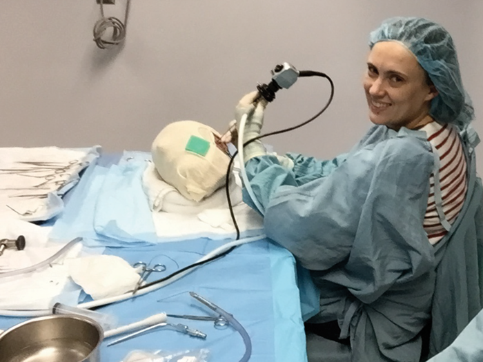
Catherine Smyth on the 5th Endoscopic Ear Surgery Course.
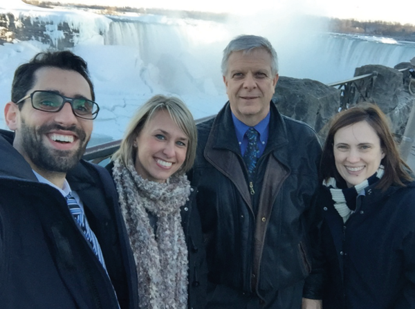
Niagara Falls with [L-R] J Nassar (Peter Munk Fellow), W Dillon (research nurse), J Rutka and C Smyth.
Summary
The TWJ Fellowship to the University Health Network Toronto has been an ever-evolving fellowship offering a broad experience in otology and neurotology. Endoscopic ear surgery has been an exciting recent addition to the programme. The learning curve is long but rewarding, with the technique offering clear advantages to the patient. Enthusiasm for minimally invasive endoscopic ear surgery is growing rapidly among ENT surgeons in Canada and globally.
Further information
http://twjfoundation.org
http://www.iwgees.org
Declaration of Competing Interests: CS has been awarded an educational stipend by the TWJ Foundation for a six month fellowship.