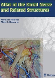
Although we all know the course of the facial nerve is pretty complex, I was surprised that a whole book and 104 pages had been dedicated to describing it. However, on reading through the book, it is apparent that although the aim of this book is to deal with the anatomy of the seventh cranial nerve, in addition, the atlas covers facial anatomy in significant detail with pictures that are clearly annotated and thorough anatomical descriptions.
The book is organised into sections: intracranial region and skull, upper facial and midface region, and lower facial and posterolateral neck region, with images that are displayed from superficial to deep dissections. The quality of the pictures are excellent and the authors use colour photographs of precisely dissected latex injected cadaver specimens, to cover the entire course of the nerve from the brainstem to its terminal branches.
Included with your purchase is access to 3D versions of all of the images and a fold-out 3D image viewing device, cleverly packaged into the back of the book. However, there are certain computer requirements to be able to use this facility and unfortunately the NHS computers that I have tried it out on were not up to speed to be able to do this!
Looking at the authors; a plastic surgeon and a neurosurgeon, this book has been printed with a facial plastic surgeon in mind and, unless this is your subspecialty area, you may not find it particularly useful. It will be beneficial for trainees wanting to understand facial anatomy in more detail or to get a grip of the complex course of the facial nerve and, although a very useful adjunct, I think I would be more likely to purchase an atlas that covers the whole of head and neck anatomy, rather than such a focused anatomy aid.


