The use of fluorescence imaging is well established in the medical sphere, forming an essential arm of medical diagnostics with liver function, ophthalmic angiography, and assessment in cell biology with fluorescence microscopy. Fluorescence imaging in surgery, however, is an evolving field, and despite over 70 years passing since its first reported use in 1947, it is still far from being universally used in routine practice.
However, with the advent of new digital cameras and technologies, real-time feedback with fluorescence is looking to change our surgical practice for the better. This article will briefly look into the current developments and future visions of fluorescence in ENT surgery using indocyanine green (ICG).
Fluorescence guided surgery
Fluorescence guided surgery (FGS) is a technique that aids in the identification of specific structures in real time that may not be normally visible under white light. A variety of fluorophores exist; however, we will consider indocyanine green (ICG) as this has gathered the greatest evidence base over time, is readily available, cheap, and is licensed for use by the US Food and Drug Administration (FDA) and UK Medicines and Healthcare products Regulatory Agency (MHRA) (although only currently licensed for diagnostic uses in liver, cardiac and ophthalmic studies, and is otherwise off-label).
“Indocyanine green is a hydrophilic fluorescent dye that is administered intravenously and binds to plasma or lymphatic proteins”
What is ICG?
Indocyanine green is a hydrophilic fluorescent dye that is administered intravenously and binds to plasma or lymphatic proteins. It absorbs near-infrared light and emits fluorescence within nanoseconds; however, this is outside of the natural visible spectrum and therefore requires detection using a camera. Timing from injection to visibility is theoretically five-to-15 seconds, and this remains in the vascular system for around five minutes before it is removed by the liver and excreted in bile without any metabolites.

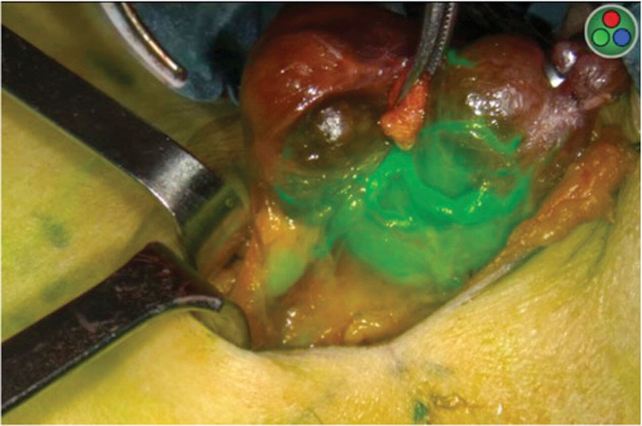
ICG being used to assess the vascularity of the parathyroid glands after thyroidectomy. Images show a very good blood supply to both parathyroid glands and therefore the patient is unlikely to have hypoparathyroidism. Top left: white light mode. Middle left: monochromatic mode displaying infrared signal. Bottom left/right: overlay of regular white light with ICG fluorescence in green. Images used with permission from Professor Frédéric Triponez.
Intraoperative injection of ICG to identify feeding vessel to aid with preservation.
Is ICG safe?
ICG is contraindicated in patients with hyperthyroidism as well as iodine hypersensitivity due to the presence of sodium iodide. Severe side effects are reported by the MHRA as very rare (<1:10,000), and include anaphylactic reactions and cardiac arrest amongst more minor ailments such as rashes and flushes.
Current benefits of ICG
One major benefit of ICG is the fact that it works under near infrared light, thereby giving greater vision into deeper lymphatic and vascular structures since overlying tissues are much more translucent in this wavelength. In addition, as opposed to normal angiography, ICG is radiation free. There is a growing number of systematic reviews suggesting the positive outcomes with ICG use across multiple specialties, such as reducing the chance of anastomotic leaks, graft necrosis, improving sentinel biopsy diagnostic rate, or use in flap assessment.
Future use of ICG
There is a growing interest in ICG usage in ENT as shown by a boom in publications over the last five years. ICG has been trialled in the aiding of diagnosis of oropharyngeal and laryngeal carcinomas, as well as in endonasal surgery for the identification of skull base tumours, and assessment of nasoseptal flap vascularity.
“One major area that is being heavily invested in is ICG angiograwphy in reducing postoperative hypoparathyroidism in thyroid and parathyroid surgery”
One major area that is being heavily invested in is ICG angiography in reducing postoperative hypoparathyroidism in thyroid and parathyroid surgery. Here, ICG is hypothesised to improve preservation of parathyroid gland vasculature and, additionally, can also assess the blood supply of the parathyroid gland by measuring the intensity of fluorescence. This may predict the gland’s function as accurately as intraoperative parathyroid hormone (PTH). Randomised trials and systematic reviews have been promising, and this may change our future practice by reducing postoperative PTH and calcium measurements and tablet supplementation.
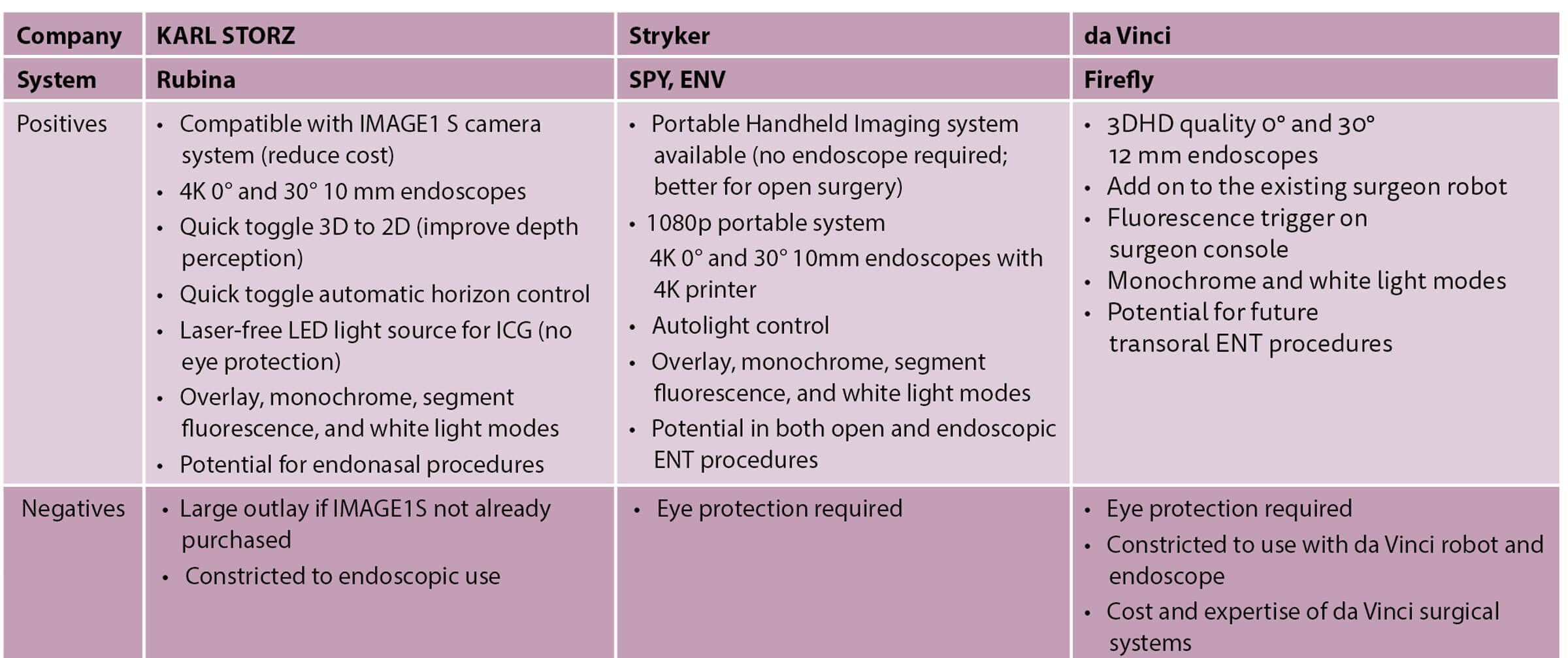
Currently, this technology is possible with hardware from multiple companies such as KARL STORZ (Rubina), Stryker (SPY, ENV; endoscopic near infrared visualisation), and da Vinci (Firefly). All of these companies use high-quality endoscopes that are capable of capturing high-definition images in white light, alongside fluorescence detecting modes. STORZ and Stryker have an additional overlay mode where ICG detection can be superimposed onto white light images. Notably, the Rubina system is compatible with existing IMAGE1 S camera systems which could reduce the overall cost of purchase. However, Stryker’s Portable Handheld Imager (PHI) allows for 1080p fluorescence imaging without an endoscope, which would be more appropriate for open surgery such as in thyroidectomy.
Conclusion
Near infrared ICG intraoperative fluorescence imaging is demonstrating exciting potential in the future of ENT, aiding advances in open and endoscopic surgeries.