In the first of a two-part series, Martyn Barnes and colleagues discuss indications for functional endoscopic sinus surgery (FESS), the surgical objectives and techniques, patient expectations and the risks of surgery. In part two, the authors will discuss how to make FESS easier, review the imaging pre and intraoperatively, optimise the operative field, decide the extent of surgery, and manage any complications.
Introduction
We have written this manuscript to highlight some of the most important things we feel we have learned in our practice of functional endoscopic sinus surgery (FESS). No short article can in itself be a substitute for experience, but we hope it provides some inspiration to our juniors, and some consolidation and reinforcement to our trainees and peers. We would welcome further dialogue to add to our thoughts.
We have tried to include points of contention by taking a stance when reasonable, and providing arguments for and against when we remain undecided. We make no apology for the ‘poor’ quality of the evidence (level III) provided in this article - it is based on our professional experience having collectively spent many years in increasingly sub-specialist rhinologic positions, learning from surgeons with a greater experience than ourselves, The techniques described have been practised and refined in theatre, and the results evaluated in the outpatient clinic.
Indications for FESS
FESS is most commonly performed in our practices for the treatment of medically refractory chronic rhinosinusitis (CRS with and without polyposis) and dental sinusitis (often unrecognised and labelled as CRS). It is also an essential surgical approach to antrochoanal polyps, epistaxis management, sinonasal tumours and skullbase surgery.
It probably has a role in recurrent acute sinusitis, although this is a much less studied (and uncommon) entity.
In most cases of inflammatory disease, FESS is indicated only after failure of medical treatments (predominantly corticosteroids and saline washes), although in many patients one can predict a poor response to medical interventions from the outset.
Surgical objectives and technique
We recommend watching our instructional video published at www.SurgTech.net/FESS/. A QR code link is provided – see Figure 1.
Surgical objectives
FESS in CRS is always performed with a number of competing objectives in mind. These include:
- Restoration of nasal patency, without excessive exposure.
- Improved delivery of medications and washes.
- Improved exposure to olfactory stimuli.
- Clearance of inflammatory foci (opacified cells and sinuses).
- Maintenance and restoration of natural mucociliary pathways.
Some of these are conflicting objectives which we must balance – optimising medication delivery (predominantly exposure of the middle meatus) may reduce olfactory exposure (the spheno-ethmoidal recess). Similarly, excessive nasal patency is reportedly associated with a syndrome of ‘empty nose’ characterised by dryness, crusting, subjective obstruction and sometimes pain. This is widely considered to relate to excessive inferior turbinate resection and is by no means an inevitable or predictable consequence. There may be important geographic and climatic factors.
Surgical techniques – principles to optimise physiology
- Know the patient’s disease extent and anatomy – see Figure 2 and ‘Imaging review’ in our next article.
- Clear polyps – use caution in polypoid change (e.g. from the turbinates and septum) which may be more vascular.
- Follow functional pathways where possible, although we do approach the posterior ethmoids through the basal lamella of the middle turbinate so as not to destabilise and lateralise it, as can happen with a more medial approach.
- Recognise and remove diseased mucosa – skullbase and orbital decompression cases are an excellent opportunity to dissect and study normal sinonasal mucosal appearances.
- Clear disease as apparent – it is not infrequent to find obvious pan-sinus mucosal thickening on dissection in theatre despite nearly normal preoperative scans.
- As a rule, when an ostium is opened, it should be opened to the maximum extent that the surrounding anatomy allows. The mucosa overlying the lamina papyracea, sphenoid and ethmoid skullbase (which should be preserved unless grossly abnormal) is often visibly less affected than the interposed (ethmoid) septations.
- Margins to establish: When incomplete (less than ‘full-house’) sinus surgery is performed, terminate the resection at points that leave the remaining natural pathways patent. For example, lower half uncinectomy, middle meatal antrostomy and (part) resection of the basal lamella of the ethmoidal bulla, or complete spheno-ethmoidectomy preserving the upper half of the uncinate lamella and some ethmoidal bulla (cut edges left well separated).
- Leave everything tidy – clear all blood and bone fragments, and lavage copiously with normal saline at the end of the procedure.
- Avoid nasal packs – they are inflammatory, and cause discomfort. If a pack seems necessary check for bleeding from the immediate distribution of the major arteries (SPA, septal, anterior ethmoid) that may be controlled with diathermy (used with great caution around the optic nerve).
- Patients must continue douching (and applying topical corticosteroids) regularly postoperatively – we recommend douching four times a day for two weeks then daily ongoing.
Surgical techniques – principles to prevent complications
- Know the patient’s anatomy, including risk factors – see ‘Imaging review’ in our next article.
- Try to identify septations then remove them – if you can see both sides, you can remove the septation (except in cases where it contains the anterior ethmoid / turbinate branches).
- Be constantly mindful of your location in the sinuses, and proximity to the lamina papyracea, skullbase (particularly medially), optic nerve, endonasal and carotid arteries.
- While dissecting the ethmoids, progress from front to back starting low and medial – identify the orbital floor in the maxillary sinus and use it as a reference to initially stay beneath.
- While dissecting the frontal recess, dissect in an anterolateral direction towards the hard bone of the frontal process of the maxilla (and away from the lateral lamella of the cribriform plate, while also remaining anterior to the orbit).
- Where diathermy is required, minimise it (low power, short bursts), avoid it near the optic nerve, and use bipolar whenever possible. Diathermy is unlikely to be necessary except where bleeding is thought to be from the SPA, septal branch, or anterior ethmoid area – other bleeding sites should be controlled with topical patties and time (Moffett’s solution or 1:1,000 adrenalin).
Surgical techniques – principles to optimise medication delivery
- Study the nose from the perspective of a nasal spray or douche (at the internal nasal valve) – can you see widely patent cavities for future medications to access?
- Consider inferior turbinate lateralisation or reduction.
- Consider middle turbinate medialisation, or even securing the turbinate to the septum by inducing synechiae (by debriding both and / or suturing).
- Perform complete surgery – topical lavages and sprays will better penetrate if the sinus cavities are open.
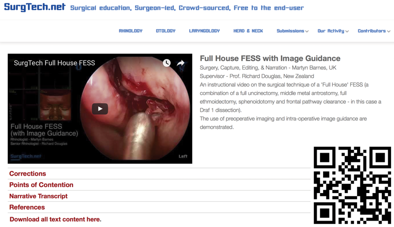
Figure 1. Learning FESS – surgical technique.
See our instructional video at www.SurgTech.net/FESS - a 12 minute clip demonstrating the surgical technique of a comprehensive FESS dissection with image guidance and radiology review. A comprehensive (or “full house”) FESS is a combination of a complete uncinectomy, middle meatal antrostomy, full ethmoidectomy, sphenoidotomy and frontal pathway clearance. If you have a QR reader, simply follow the QR code link.
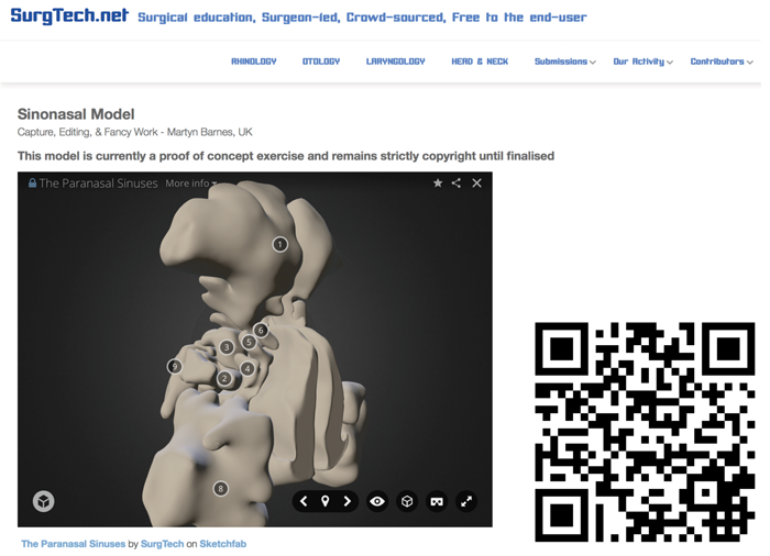
Figure 2. Learning FESS – anatomy.
We recommend learning the complicated surgical anatomy of FESS from different perspectives:
1. The endonasal view (endonasal examination and observed operating)
2. The 3-axis imaging view (operative scan review)
3. Our 3D anatomical model (an outside-in perspective that we think is much simpler).
See our model at www.SurgTech.net/sinuses. If you have a QR reader, simply follow the QR code link.
Surgical techniques – principles to optimise olfaction
- Preserve normal olfactory mucosal – the high septum, the whole of the superior turbinate, the medial aspects of the middle turbinate.
- Polyps (pedicled) should never be retained, although limited polypoid change in an olfactory region should be preserved and may recover.
- Optimise ‘exposure’ of olfactory mucosa (contrary to the approach to optimise medication delivery) and do not medialise the middle turbinate. However, unless the disease is controlled olfaction is unlikely to improve.
Patient expectations
It is critical that any patient undergoing FESS surgery (and their surgeon!) understands…
- The surgery is not curative, nor is it an alternative to medical therapy.
FESS surgery is an adjunct to medical therapy – it will usually clear the nose of obstruction, but in the vast majority of cases ongoing medical therapies are required to maintain control of both inflammation and symptoms. Surgery not only improves symptomatic control (mainly by resolving obstruction) but also exposes the sinonasal cavities to facilitate more effective medical treatment. It helps if patients consider their disease analogous to asthma – a (closely related) pathology of uncertain and variable origin, that is incurable, but certainly can be better controlled. This is one of the ways to convey the important message that they must continue their medical treatments. We usually recommend intranasal corticosteroids and saline washes lifelong until proven otherwise. - The best outcomes are rarely a complete return to ‘normal’.
Depending on the case, FESS surgery may improve nasal obstruction, rhinorrhoea, facial pressure, pain, anosmia and eustachian dysfunction. No improvements are assured but we emphasise that we are most optimistic to improve nasal obstruction. Rhinorrhoea and pressure are likely to improve, but pain and anosmia are often diagnostic uncertainties and there is unfortunately a small potential to make these symptoms worse with surgery. - There is a recovery period.
There are usually three phases of recovery – a one to two week period of nasal congestion and bleeding followed by some months of symptomatic changes dependent on the balance of the longer-term healing process, disease recurrence, and medical control. The outcome of this determines a final symptomatic plateau, long-term symptoms and in some cases the need for further surgery. - There are risks.
Patients have great difficulties weighing up the risks and benefits of the decision for surgery. Some will not want to be fully informed (we are still obliged to inform them), others will be unable to comprehend the issues, and absolutely none of them will leave our relatively brief clinic interaction with the full understanding we would like to provide. We can only do our best. Printed materials for patient education may help and are certainly recommended (we will provide open access documents at www.SurgTech.net soon and are enrolling contributors). These should describe the surgical indications, procedure, risks and complications. Such materials should be available well before the operation for the patients to consider and return with any questions.
“We have tried to include points of contention by taking a stance when reasonable, and providing arguments for and against when we remain undecided.”
Risks of surgery
FESS is commonly combined with septoplasty and turbinate surgery and the decision may not be made until the case is in progress. We therefore often consent a patient in the expectation we may be performing a comprehensive FESS, septoplasty and turbinate reductions.
Although comprehensive FESS is more likely to result in complications, the most limited sinus surgery (an uncinectomy) is the most common cause of an orbital breach, and rarely may result in a cerebrospinal fluid leak also. The following risks are therefore best discussed in all cases.
Symptomatic failure
Chronic rhinosinusitis is not cured by FESS or any other means. Although some may achieve excellent symptomatic control, many have ongoing but reduced symptoms and a small proportion will have only transient benefit (although most of these do eventually improve with medical therapy and sometimes revision surgery).
Antrochoanal polyps and dental sinusitis may have better outcomes; the latter especially when combined with dental treatment.
Ongoing treatment
All patients are informed that they will need ongoing medical treatment – usually intranasal corticosteroids and large volume saline douching (four times daily for two weeks then once daily ongoing). They should be aware that further surgery may be required.
Revision surgery is most commonly required in those patients with CRS without polyps predisposed by immunodeficiency and ciliary disorders, and in nasal polyposis cases managed too conservatively, especially in the presence of allergic fungal sinusitis.
Bleeding
All our patients should expect some postoperative bleeding, but mostly this is self limiting. Advice should be provided on first aid measures such as saline douching (warmed if necessary), sucking ice cubes, and if required, presentation to emergency services.
Intraoperative bleeding occasionally requires surgery to be discontinued, or measures including nasal packing, cautery or sphenopalatine artery ligation. In rare cases, a blood transfusion is required.
Anticoagulant use will exacerbate bleeding, but perioperative withdrawal or reversal may not always be required and should be considered on a patient by patient basis.
It is usually possible to perform FESS safely in patients on low dose aspirin. In patients with significant coronary artery disease or with cardiac stents it is generally safer to operate without withdrawing this medication. Consulting the patient’s cardiologist is advised. In contrast, newer agents such as ticlopidine are very effective anti-platelet agents and sinus surgery should generally not be performed if these drugs cannot be safely withdrawn.
Orbital injury
Orbital complications may result from any breach of the orbital periosteum (beyond the bony lamina papyracea) or from ethmoid artery injury. Small breaches are not rare, but when they are complicated by intra-orbital bleeding or in more extensive injuries, diplopia, retrobulbar haematoma, or blindness may occur. These risks are thought to be less than 1:1,000.
Skullbase injury
Intracranial complications and cerebrospinal fluid (CSF) leaks usually result from penetration of the cribriform plate and associated dura, often close to the anterior ethmoid artery. It may be recognised during the procedure, or present with clear rhinorrhoea postoperatively. The risk of such skullbase injury during FESS is thought to be less than 1:1,000.
Lacrimal injury
Lacrimal injuries are often unrecognised and many result in little symptomatic consequence. However, there is a risk of secondary epiphora, which may be managed by dacryocystorhinostomy.
Packs, splints
We avoid packs where possible, so they are infrequent, but the patient should be aware that they may require one if troublesome intraoperative bleeding is experienced. Similarly, we occasionally use nasal splints in FESS surgery.
Adhesions
Synechiae following surgery are common in the middle meatus, or between the septum and inferior turbinates. The latter may compromise early airway results and medication delivery but can usually be divided in clinic postoperatively. Middle meatal adhesions may prevent effective medication delivery and compromise longer term results, requiring (usually minor) revision surgery.
Changes to the sense of smell
In many cases, sinus surgery is performed in patients lacking a sense of smell and this can not be made any worse. In other cases, there are small risks of dysosmia, hyposmia or anosmia.
Infection
Postoperative infection may be a problem in cases of orbital breach, septal haematoma, or compromised sinus drainage. The latter (essentially acute sinusitis) is most common after frontal recess surgery, and can usually be managed with analgesia and (in cases with pyrexia or mucopurulent discharge) antibiotics. Such episodes may be the consequence of suboptimal surgical results which will also compromise longer term disease and symptom control.
Additional complications of septoplasty
Where a septoplasty is required, there are additional risks of septal perforation and collapse (leading to airway compromise and occasional saddle deformity). The septal L-strut should always be preserved or reconstituted, so these are usually the consequence of an untreated and infected septal haematoma. Numbness of the upper incisor teeth is a relatively common complication of septoplasty, but is almost always transient.
“FESS surgery in CRS is always performed with a number of competing objectives in mind.”
Additional complications of turbinate surgery
Turbinate surgery most significantly adds to the risk of bleeding, but cautious technique and attention to intraoperative haemostasis should prevent this. Depending on the technique used, postoperative crusting can be extensive and require debridement in clinic after two weeks.
There is also the contentious issue of empty nose syndrome and / or atrophic changes, largely attributed to excessive turbinate resections. In our UK experience to date, we have performed conservative resections in most cases and have never had problems with these issues. We know of centres in much drier climates that have reported no problems with much more extensive resections.
Turbinate surgery should not be dismissed in selected cases. It has the potential to facilitate medication delivery and can contribute greatly to refractory nasal obstruction. We have even seen (non-polyp) patients return delighted with the side-effect of having ‘cured’ their snoring. Nonetheless, it is not something to be undertaken without due consideration.
‘High risk’ cases
The highest risk cases are those in which the surgeon’s view is compromised by the disease, bleeding, or available equipment. Revision surgery is more challenging due to deranged anatomy, prior complications (e.g. orbital breach), and scar tissue or changes in the disease vascularity.
Conclusion
We set out to highlight some of the most important things we feel we have learned to date in our practice of FESS. We hope you have found this useful. We found so much to say that we have separated this into two parts, and will publish part two later this year, discussing how to make FESS easier, review the imaging pre- and intraoperatively, optimise the operative field, decide the extent of surgery, and manage any complications. We do invite you to comment on the article (email: martyn@surgtech.net) and to contribute to our dynamic library of this and other works at www.SurgTech.net.
Declaration of Competing Interests: This article has been contributed to by SurgTech – a non-profit, open membership organisation established to promote open access surgical education. Our members are in no way financially reimbursed for their contributions.