Brain imaging advances are revealing tinnitus-related neural networks, shedding light on its pathophysiology, heterogeneity and potential therapeutic targets.
Tinnitus is a highly common but poorly understood disorder [1]. Its subjective nature and the fact that its treatment may span multiple disciplines (such as audiology, otolaryngology, clinical psychology, physical therapy) can create uncertainty for both patients and healthcare professionals.
Another reason for the elusive nature of tinnitus is a limited understanding of its mechanisms and how they relate to the symptoms and challenges faced by patients. The lack of definite knowledge about its mechanisms in humans leads to poorer subtyping and less favourable treatment outcomes.
Over the past two decades, advances in neuroimaging have fundamentally altered our understanding of tinnitus. Once considered primarily a consequence of abnormal activity within the auditory periphery, tinnitus is now recognised as a disorder involving distributed brain networks that influence attention, emotion, memory and perception. These discoveries have reshaped current theories of tinnitus generation and persistence, while providing new opportunities for patient stratification and treatment development. The tools used vary in terms of spatial and temporal resolution as well as the experimental paradigms. Advances in structural MRI, functional MRI, diffusion imaging and electrophysiological techniques have enabled researchers to investigate both the anatomy and function of tinnitus-related brain networks.
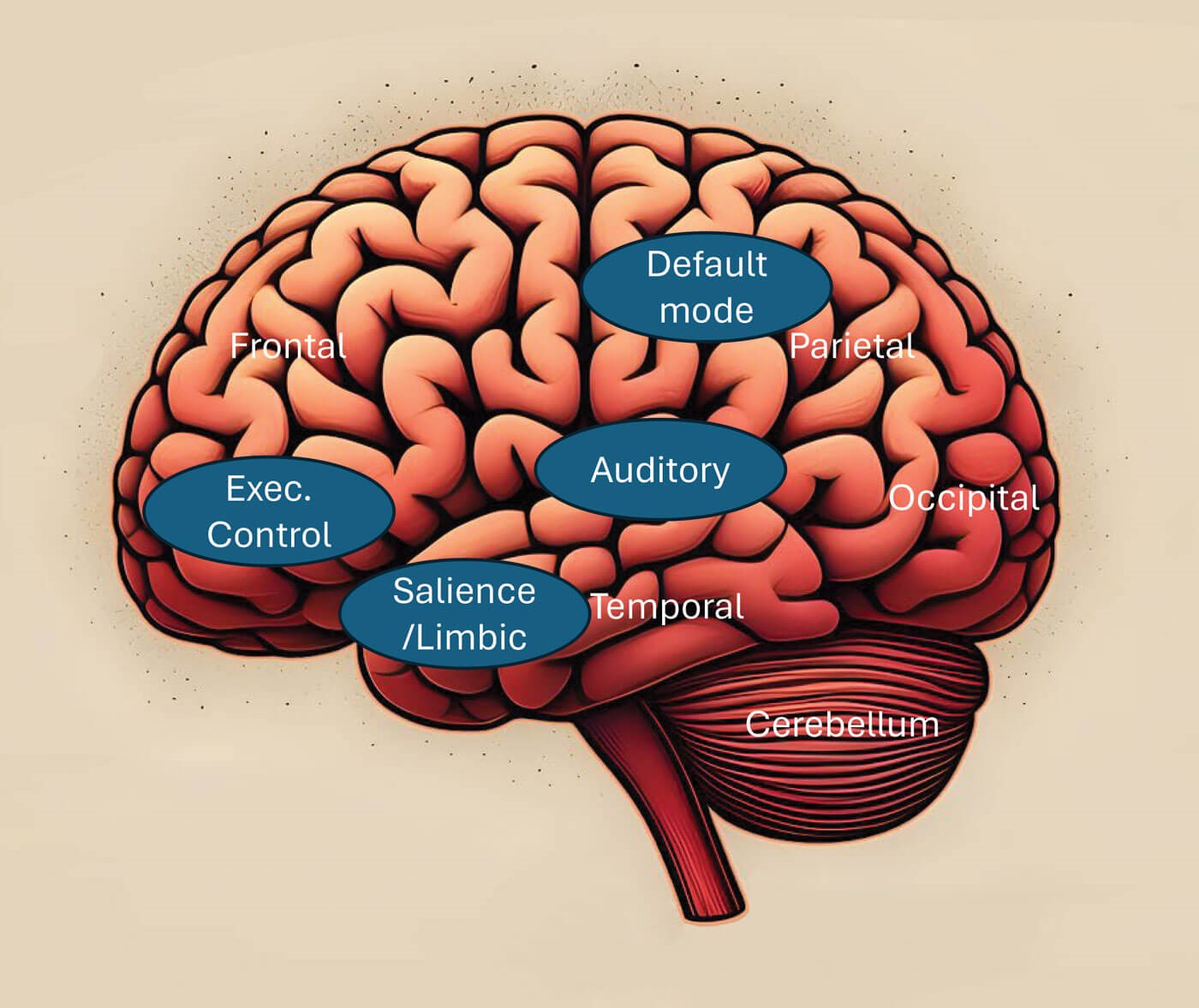
Figure 1: The typical tinnitus-related networks in the brain. Actual nodes of the networks are noted in the table.
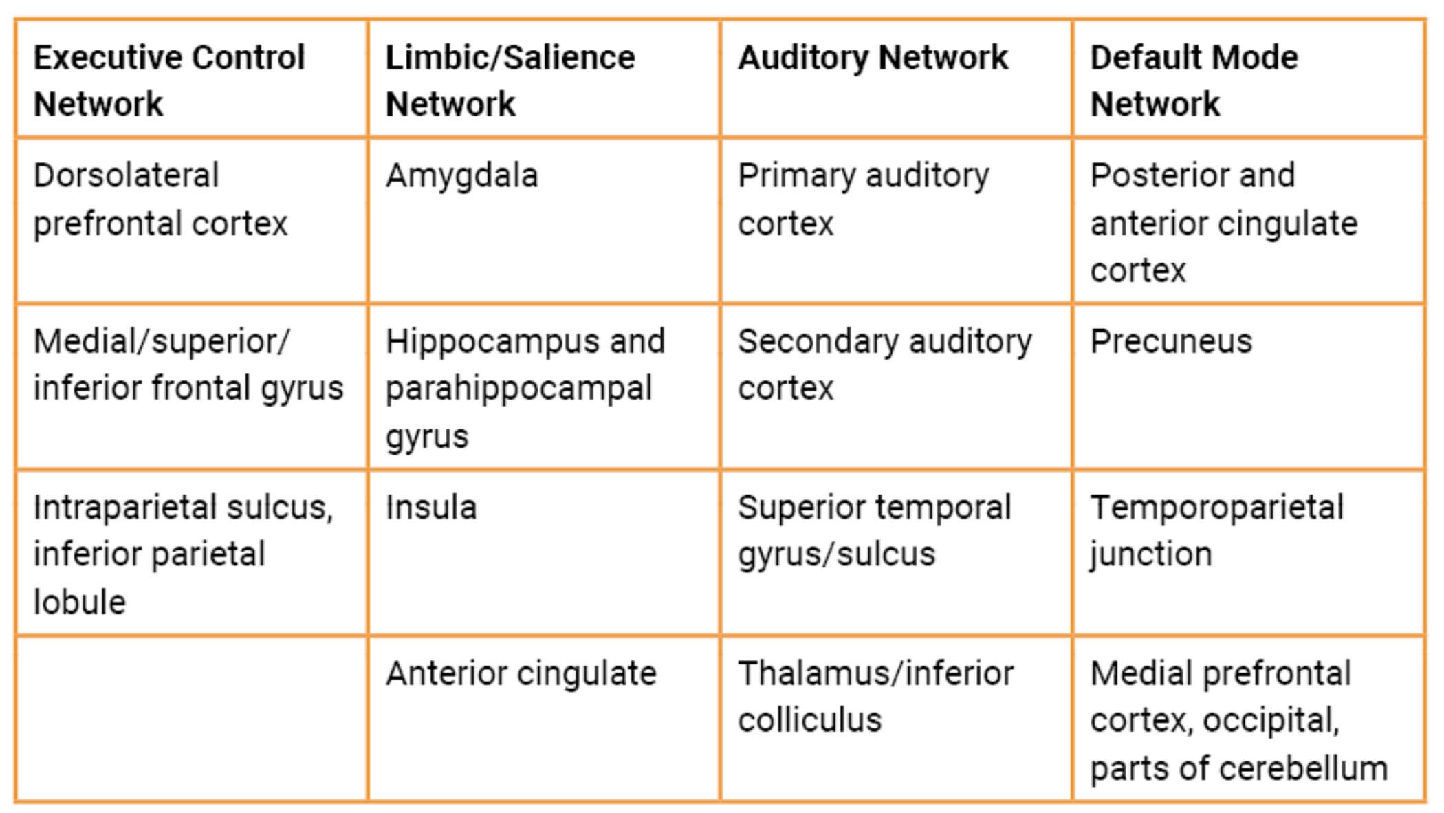
Figure 1 and the associated table depict the major neural networks associated with tinnitus (for more details see Husain and Khan [2] and Shahsavarani et al. [3]). The executive control network is primarily concerned with top-down regulation of attention and interacts with the limbic system to regulate emotions. Emotion processing is primarily mediated by the limbic system and the salience network. The auditory network processes sound and comprises the primary and secondary auditory cortices, along with the central auditory pathways. The default mode network is most active during rest or periods of quiet, and supports self-referential thinking, autobiographical memory, future planning and conscious awareness of sounds.
"Once considered primarily a consequence of abnormal activity within the auditory periphery, tinnitus is now recognised as a disorder involving distributed brain networks"
Major findings and applications of neuroimaging
This section reviews current knowledge derived from extensive evidence that was unavailable two decades ago, highlighting key findings and applications of neuroimaging.
- Periphery vs. cortex: Evidence from neuroimaging suggests that peripheral mechanisms, including hearing loss and abnormalities of middle and inner ear function, are unlikely to fully account for the generation and persistence of tinnitus. Although hearing loss remains the strongest known risk factor, imaging studies indicate that tinnitus is also associated with alterations in distributed central neural networks.
- One site vs. networks: Earlier studies were focused on one brain region, such as the dorsal cochlear nucleus or inferior colliculus as the site for the generation of tinnitus. With neuroimaging, unlike animal studies, we can obtain information from all regions in the brain simultaneously. Increased involvement of salience, emotion-processing (limbic) and attention networks may explain why some individuals are unable to ignore tinnitus, despite similar hearing loss to those who experience little distress.
- Heterogeneity: Brain imaging is able to provide information about the neural bases of different subgroups, varying on the basis of symptom severity or the existence of comorbid conditions such as hearing loss, hyperacusis or posttraumatic stress disorder. In particular, the strengths of the functional connections between the tinnitus-related networks may differ based on symptom severity or comorbid conditions.
- Evaluating treatment effects: In recent years, longitudinal studies have mapped out treatment effects of the most commonly used treatments, such as hearing aids and cognitive behavioural therapies. For instance, better emotional regulation as a result of mindfulness-based cognitive therapy, as noted in tinnitus-related questionnaires, can be linked to specific functional changes in the default mode, executive control and limbic networks [4].
- Imaging-guided treatments: Neuroimaging has identified novel therapeutic targets, particularly within non-auditory brain networks involved in attention and emotional processing. Imaging-guided neuromodulation approaches, including transcranial magnetic stimulation and transcranial direct current stimulation, have shown encouraging results in some studies, although evidence remains insufficient for routine clinical implementation.
Based on the responsiveness of neuroimaging to the inherent variability of the patient population and to changes due to treatment, the neural correlates identified by these tools can serve as biomarkers for the subjective condition, rather than simply as outcome measures.
"Brain-imaging-guided treatments... have shown some promise but not consistently enough to be deployed in clinics"
Presently, one limitation of brain imaging studies is the small sample sizes. Big data, including data from multiple centres and studies, along with advances in analytical methods, namely machine learning and artificial intelligence, provide one solution moving forward. Combining data also has the potential for finding invariant signatures of tinnitus while also allowing for better subtyping.
Future priorities in using neuroimaging include the development of large international imaging consortia, multimodal imaging datasets and clinically useful biomarkers. Integrating neuroimaging with complementary approaches such as genomics, electrophysiology and digital phenotyping may ultimately support a precision-medicine approach to tinnitus, in which treatments can be tailored to the biological profile of individual patients.
References
1. Vanneste S, De Ridder D, Gallus S, et al. Tinnitus. Nat Rev Dis Prim 2026;12(1):29.
2. Husain FT, Khan RA. Review and Perspective on Brain Bases of Tinnitus. J Assoc Res Otolaryngol 2023;24(6):549–62.
3. Shahsavarani S, Khan RA, Husain FT. Tinnitus and the brain: a review of functional and anatomical magnetic resonance imaging studies. Persp ASHA Spec Inter Sig 2019;4(5):896–909.
4. Zimmerman B, Finnegan M, Paul S, et al. Functional brain changes during mindfulness-based cognitive therapy associated with tinnitus severity. Front Neurosci 2019;13:747.
Declaration of competing interests: None declared.