In his second article on this topic (see here for the first article), Richard E Gans explains how to use vestibular rehabilitation therapy to treat vestibular patients, and demonstrates why this method of diagnosis based strategies has proved so successful.
Introduction
Common inner ear disorders that cause vestibular dysfunction include: labyrinthitis, vestibular neuritis, herpes zoster oticus, vestibular migraine, labyrinthine ischemia and Ménière’s disease. Most patients may have had only a relatively short phase of acute vertigo, or are post-surgery for treatment of intractable inner ear disease such as Ménière’s disease.
Once out of the acute phase they may be left with chronic symptoms affecting their sense of spatial orientation, gaze stabilisation during activities, or balance. This classification of post-acute but symptomatic patients are considered to be experiencing a stabilised but noncompensated vestibulopathy. It is these patients who are the ideal candidates for vestibular rehabilitation therapy (VRT). VRT works best when it is used with individuals who are outside of the acute phase of a condition [1].
Physiological basis of VRT and central compensation
VRT consists of systematic repetitive exercises and protocols that extinguish, or ameliorate patients’ motion-provoked symptoms, reset the gain or precision of the vestibular ocular reflex (VOR) as well as enhancing postural stability and equilibrium. It allows the brain to see the error signals coming from the impaired labyrinth. The underlying physiological basis for VRT is the plasticity of the central nervous system. Possible mechanisms include the spontaneous rebalancing of the tonic activity within the vestibular nuclei, recovery of the VOR through adaptation, and the habituation effect, that is a lessening of response to the same stimuli over time. In addition, the flocculus within the cerebellum and more recent research with PET scans indicating additional compensation within the cortex has been published [2].
“The underlying physiological basis for VRT is the plasticity of the central nervous system.”
Theoretically, central compensation should occur within 90 days following dysfunction or loss of one of the vestibular systems. Central compensation may be delayed or ineffective due to the reluctance of patients to perform activities that produce symptoms of dizziness. Other complicating factors include commonly prescribed pharmaceuticals that suppress either peripheral vestibular or CNS function. These drugs will delay or prevent the central nervous system from relearning or adapting to asymmetrical sensory input. Unfortunately, the dizzy patient, in his / her heightened state of anxiety about becoming dizzy (especially while at work or driving), becomes reliant on those pharmaceuticals.
“Diagnosis based strategies is an individualised or customised therapeutic VRT approach which has been shown to produce successful outcomes.”
Abbreviation Key
m-Hallpike - Modified Hallpike, BPPV - Benign Paroxysmal Positional Vertigo, DVA - Dynamic Visual Acuity, PC - Posterior Canal, HC - Horizontal Canal, SOP - Sensory Organization Performance test, DGI - Dynamic Gait Index, CTSIB - Clinical test of Sensory Integration of Balance, CRM - Canalith Repositioning Manuever, VOR- vestibular ocular reflex.
Diagnosis-based strategies: the brain cannot fix what it cannot see
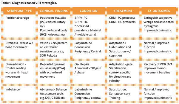
Diagnosis based strategies is an individualised or customised therapeutic VRT approach which has been shown to produce successful outcomes [3]. These strategies link the underlying physiological changes that occurred due to the disease or insult with the patient’s functional symptoms. Table 1 provides an overview of common symptoms and the corresponding theraputic protocols. There are several approaches to therapy [4]:
- Adaptation will reset or retune the VOR by repetitive activities. These activities will include those situations or movements that provoke the very symptoms the patient has been trying to avoid. Examples of the excercises are shown in Figure 1. A complex activity will incorporate gaze stabilisation exercises; mostly coordinated head and eye movement along with head and eye movement with either full ambulation under dynamic conditions. Presenting the patient with multiple simultaneous acceleration stimuli in a variety of planes will provide an excellent activity with a minimum of space, cost and equipment. A good example of this would be to have the patient sit on a balance ball, with a slight bounce while they turn their head from side-to-side and read two separate word lists, like a grocery list. Baseline, serial or final performance may be evaluated with any test of dynamic visual acuity [5].
- Habituation exercises, as shown in Figure 2, reduce the hallucination of motion or movement as well as extinguishing the sensation of after-motion. This too, is based on neural plasticity and works only through the systematic repetition of the movements, acceleration with speed or direction that provoke the symptoms. The brain is exposed to the noxious stimuli repeatedly in a short time span. The patient’s subjective report of the intensity of the motion and duration of after-motion are utilised to determine treatment efficacy.
- Substitution protocols, as shown in Figure 3, will strengthen the weakened systems by reducing the dependence on the remaining ones. Or, in the case where a sensory modality is missing, will work to strengthen or make the remaining systems more trustworthy. A patient with a weakened vestibular system is forced to make it more dominant by reducing or challenging the somatosensory input by standing on a trampoline. The visual sense could be further disrupted or diminished by having the patient close his eyes or watch a moving visual stimulus while maintaining his balance.
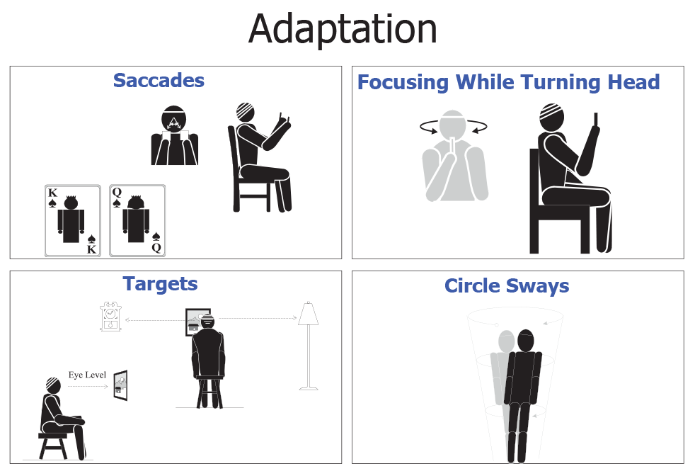
Figure 1: Adaptation [4].
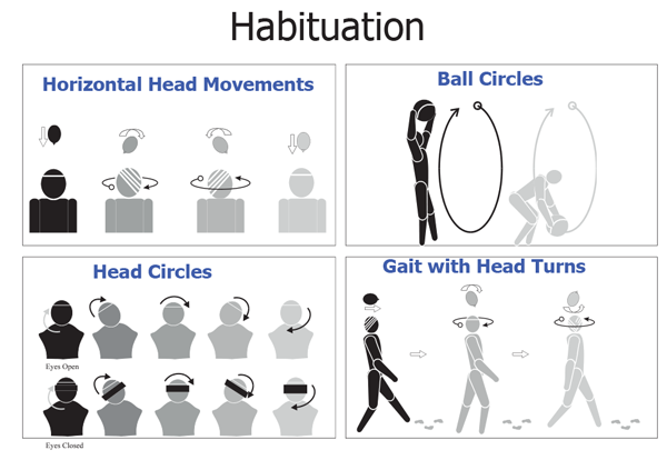
Figure 2: Habituation [4].
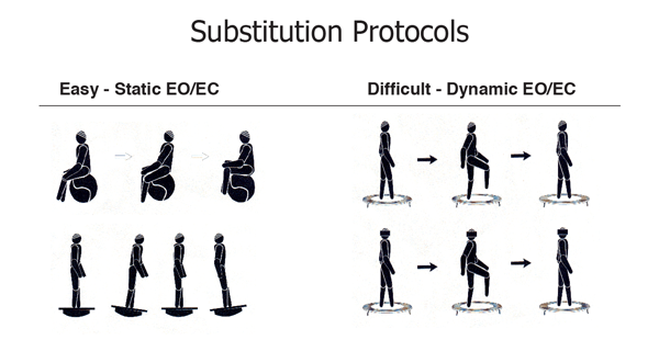
Figure 3: Substitution protocols.
“It is important that the patient’s clinic sessions be supplemented by at-home activities, as the one to three hours per week in clinic will be insufficient to provide an efficient and efficacious outcome alone.”
Typically VRT will be conducted in clinic at least one to three times per week for three to five weeks, each session lasting 45-60 minutes. Most patients will do very well and be ready for discharge within 30-45 days. It is important that the patient’s clinic sessions be supplemented by at-home activities, as the one to three hours per week in clinic will be insufficient to provide an efficient and efficacious outcome alone. At-home sessions should be performed by the patient daily for at least 15-30 minutes, and for those who can perform two or three 15-20 minutes sessions, that would be ideal. Summary Since the initial publications of Cawthorne and Cooksey over 60 years ago, hundreds of research articles, and succesful outcomes with tens of thousands of patients worldwide have clearly solidified VRT as the gold standard for the non-medical treatment of non-compensated vestibular disorders. Patients who suffered with chronic non-acute, but debilitating symptoms, are no longer told to “learn to live with it” but can recover their ability to return to normal everyday activities of work and play.
References
1. Gans, RE. Vestibular rehabilitation therapy. In: Dispenza F, De Stefano A, (Eds.). Textbook of Vertigo Diagnosis and Management. London, UK; Jaypee Brothers Medical Publishers; 2013.
2. Gans RE. Vestibular Rehabilitative Therapy. In Katz, J, (Ed.). Handbook of Clinical Audiology 7th Edition. Philadelphia, PA, USA; Wolters, Kluwer Health; 2015. 3. Black FO, Angel CR, PeszneckerSC, Gianna C. Outcome analysis of individualzed vestibular rehabilitation protocols. Am J Otol 2000;21:543-51.
4. Gans RE. Vestibular Rehabilitation: Protocols and Programs. Tampa, FL, USA; AIB Education Foundation Press; 2010.
5. Roberts RA, Gans RE. Comparison of horozontal and verical dynamic visual acuity in patients with vestibular dysfunction and nonvestibular dizziness. J Am Acad Audiol 2007;18:236-44.
Declaration of competing interests: None declared.