
The ENT team has always been involved in securing long-term enteral feeding in complicated cases. This technique offers a novel approach for the specialty to achieve this.
Passing an NG or Ryle’s tube is a blind procedure. The tube can traumatise the nasal mucosa, coil in the pharynx, enter the trachea or bronchial tree, perforate the oesophagus, or, in the presence of a skull-base injury, pass intracranially. Across almost 10,000 blind narrow-bore placements, tracheobronchial malposition occurred in about 1.9%, pneumothorax in about 0.35%, and death in about 0.05% [1].
Delivering feed or medication through a tube that is lying in the lung or pleura is classified in the United Kingdom as a ‘Never Event’, judged wholly preventable when existing guidance is followed. The National Patient Safety Agency recorded 21 deaths and 79 cases of harm from this event between 2005 and 2010, and national bodies have reissued placement guidance more than once since [2,3]. Harm has continued despite these alerts, partly because the two bedside confirmation methods – pH testing of aspirate and radiograph interpretation – can each fail [4].
Replacement carries the same risks as the first passage, and often more. A blocked, displaced or malfunctioning tube must be exchanged, and malposition has been reported in 13% to 32% of repositioning attempts [1]. When passage is difficult, described adjuncts include stiffening or chilling the tube, positioning and rotation manoeuvres, flexible nasendoscopy, video laryngoscopy and guidewire assistance [5–7].
These methods address the initial passage. Less attention has been given to preserving and reusing the tract a functioning tube has already established, which is the specific problem the technique described here addresses.
Technique
The technique was performed on a rigid sagittal anatomical model of the head, neck and upper gastrointestinal tract. Each step below refers to that setting.
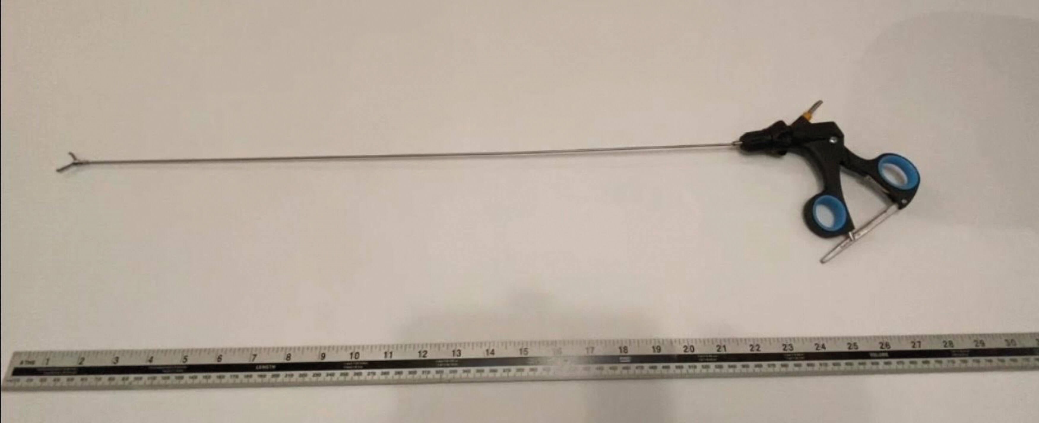
Figure 1: A 60-cm-long, slender forceps with a locking mechanism and toothed jaws.
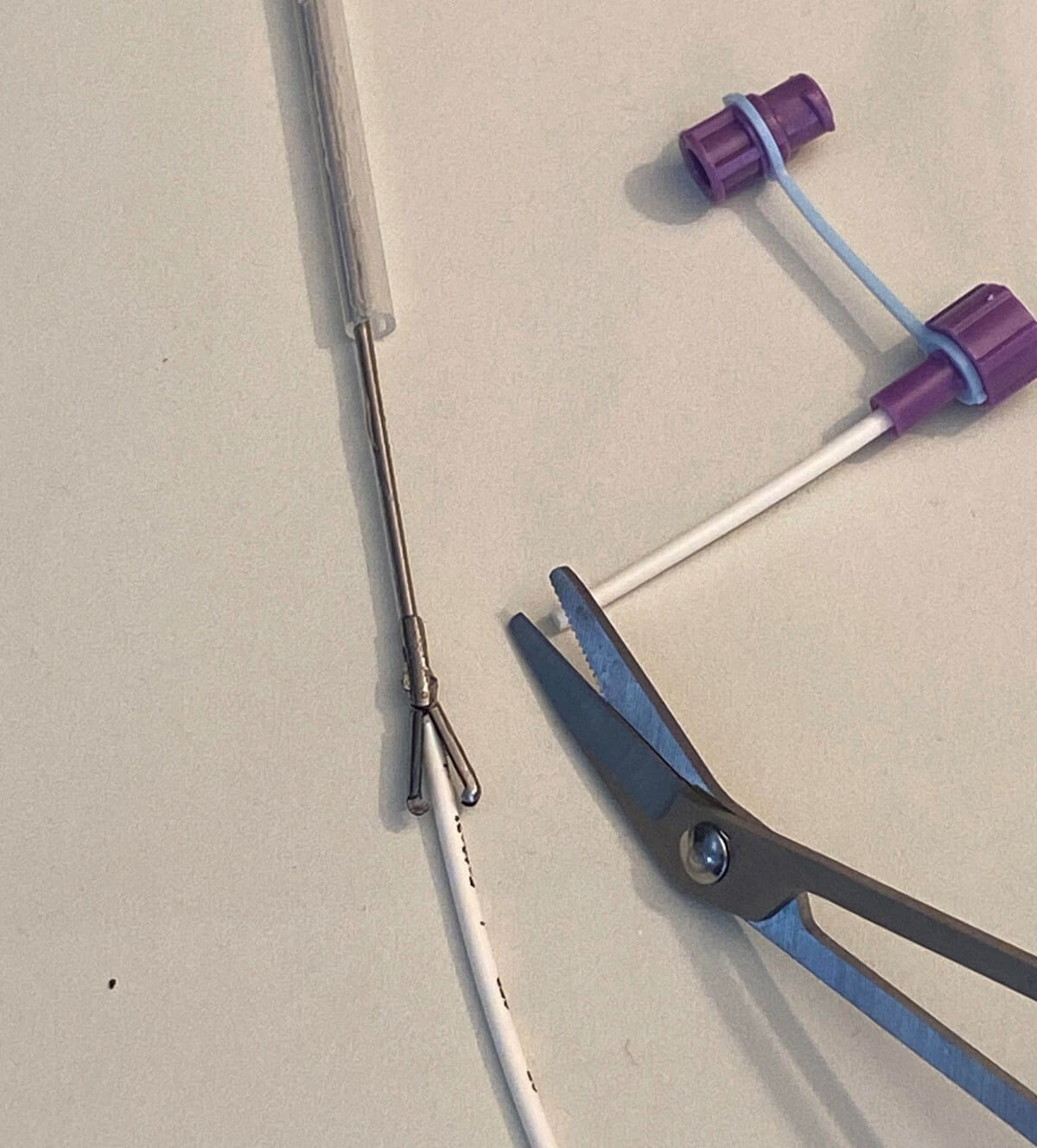
Figure 2: The lubricated silicone conduit threaded over the long forceps,
with the trimmed proximal end of the existing NG tube held securely.
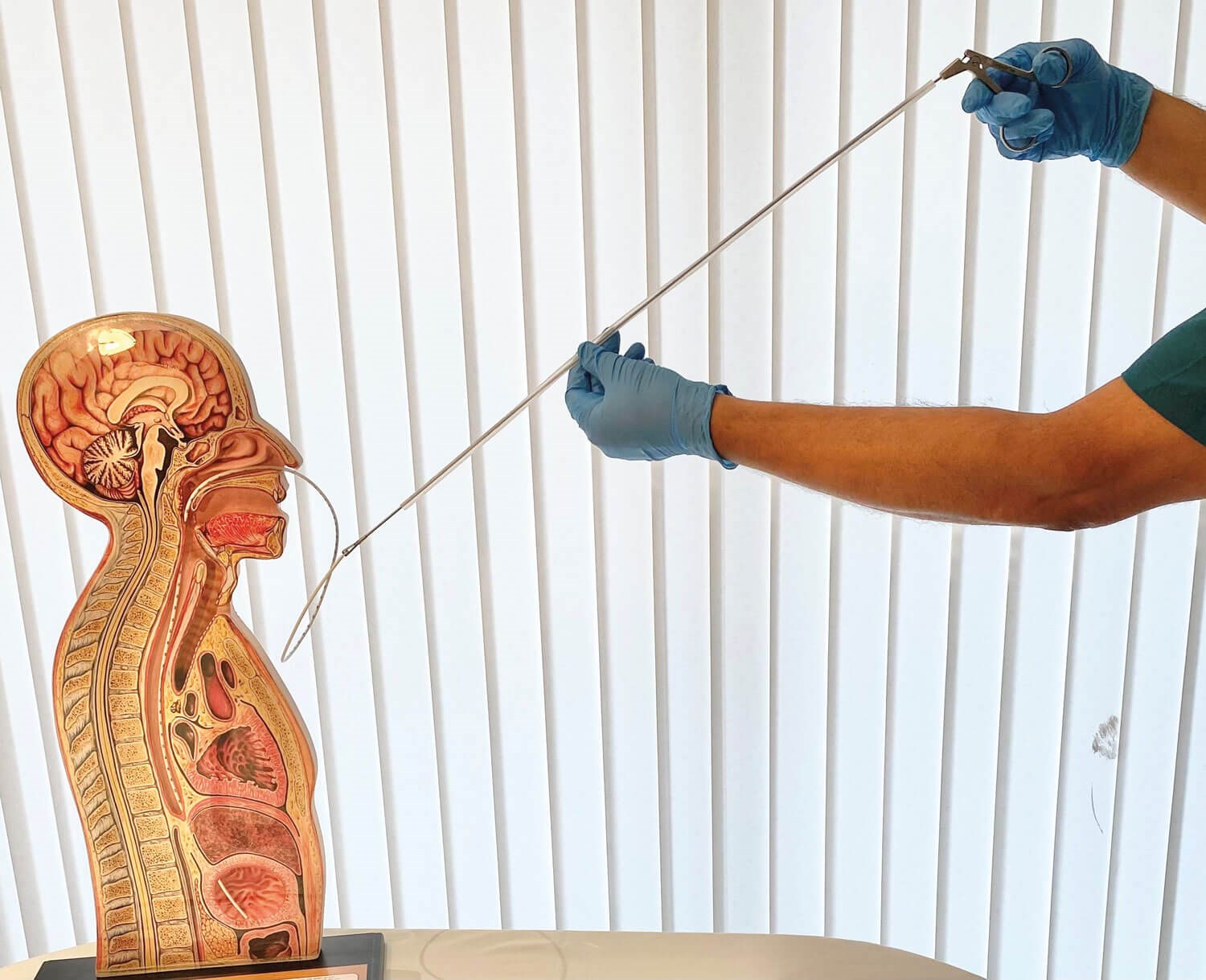
Figure 3: The silicone conduit is gradually advanced through the nose and into the stomach, over the existing NG tube.
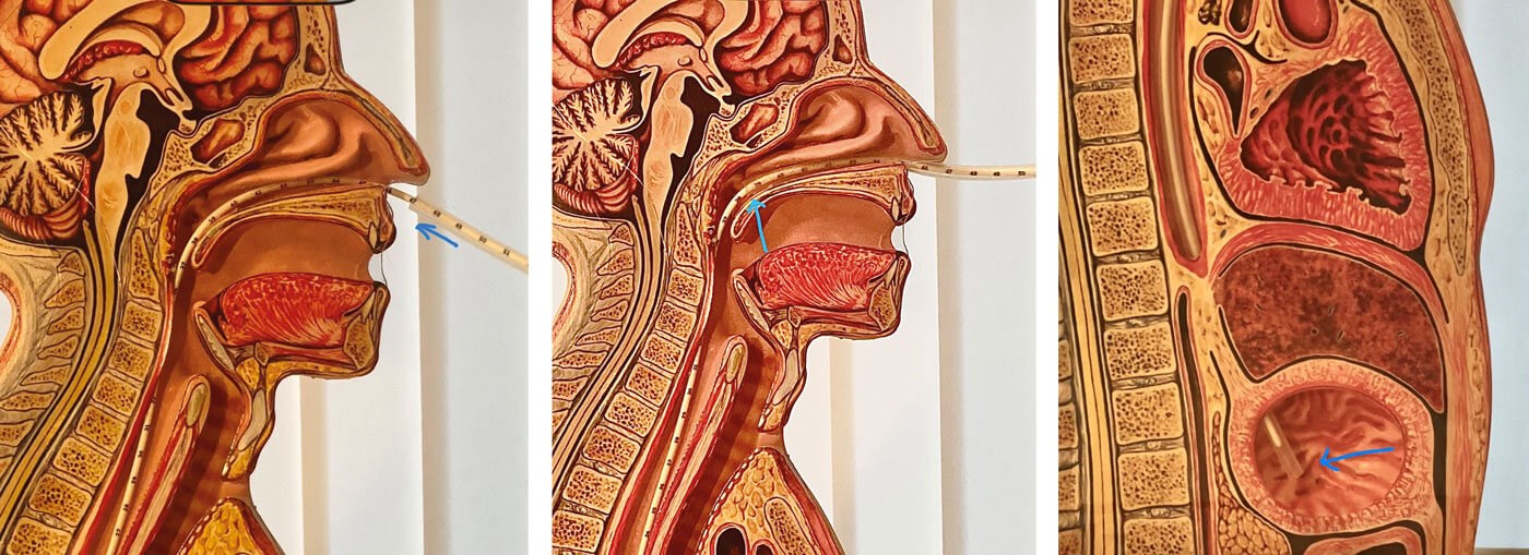
Figure 4a–c: The silicone conduit is advanced through the nose, over the existing NG tube, and into the stomach.
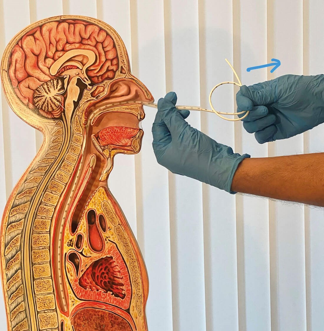
Figure 5: The old NG tube is withdrawn through the silicone conduit.
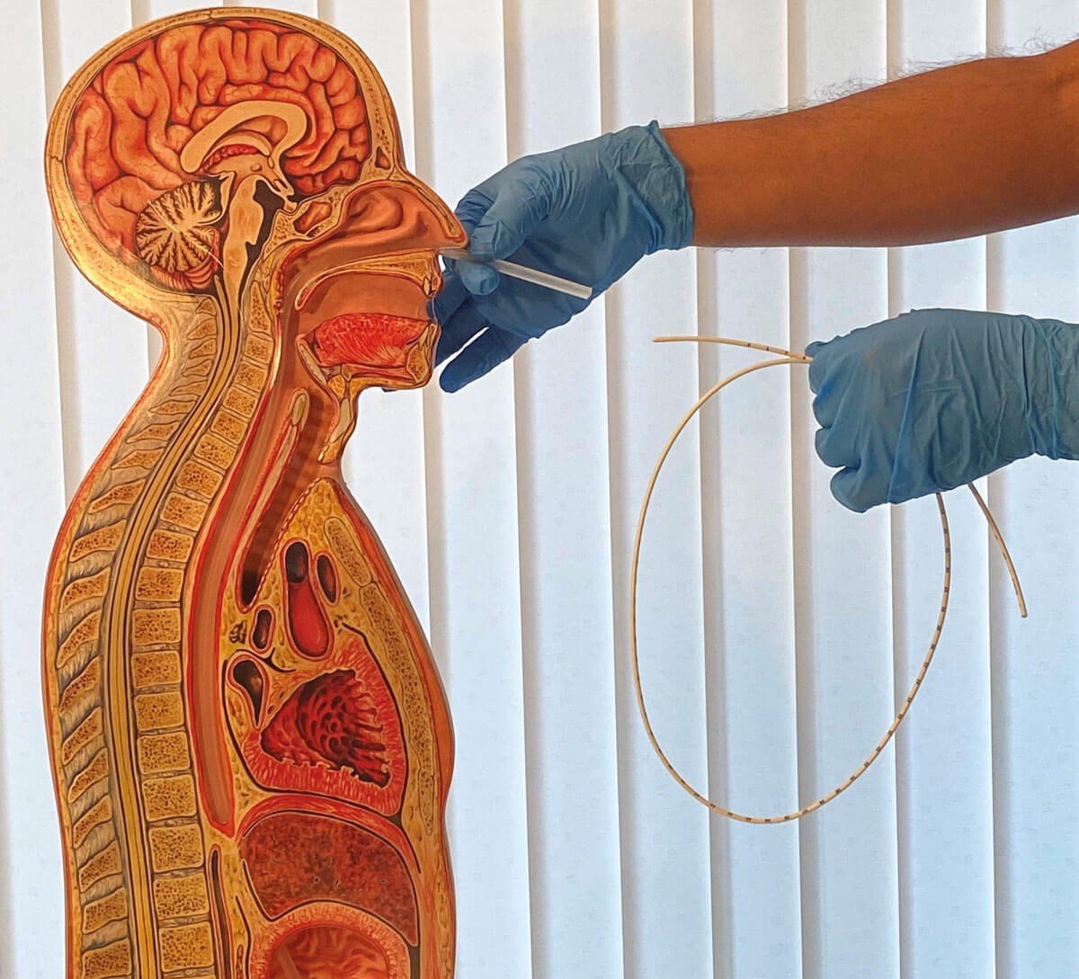
Figure 6: The empty silicone conduit remains in place from the nose to the stomach after removal of the old NG tube.
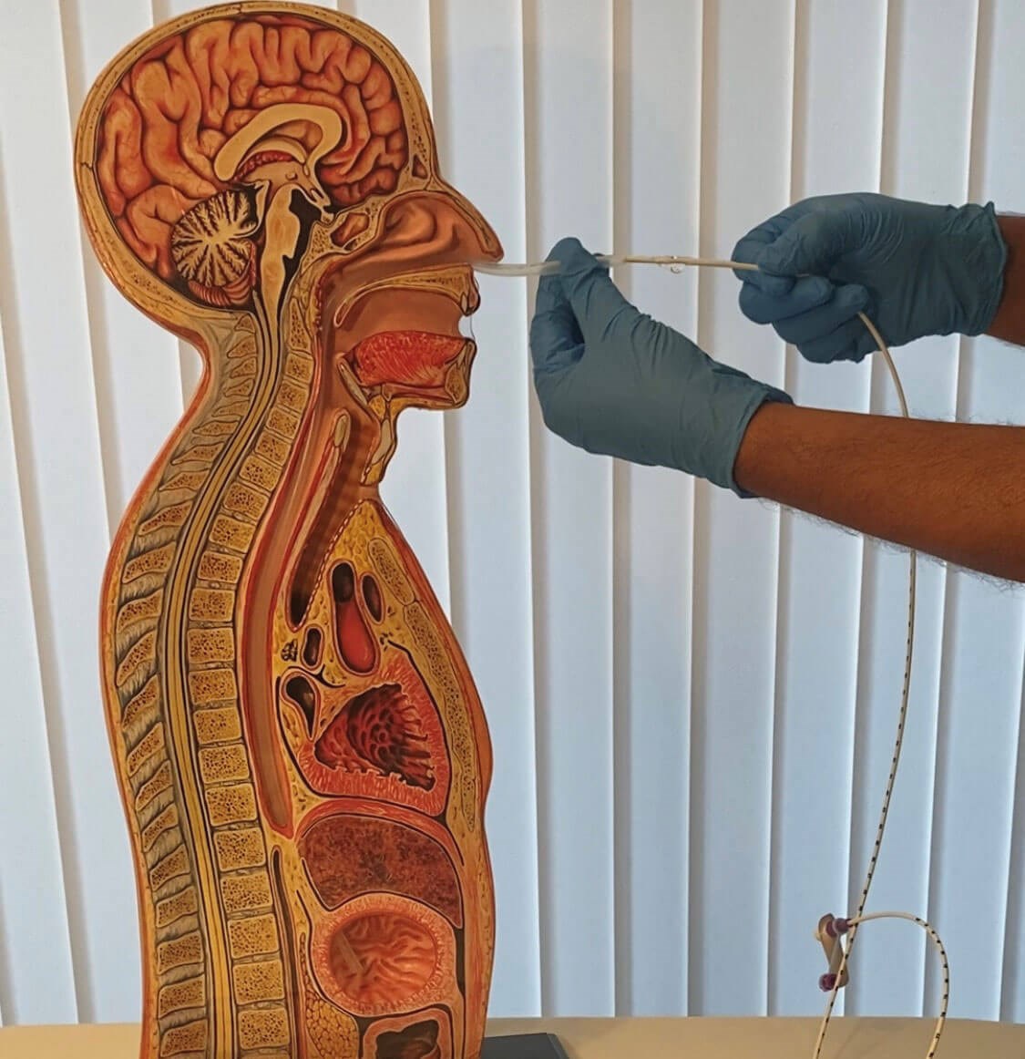
Figure 7: Insertion of the lubricated new NG tube through the silicone conduit.
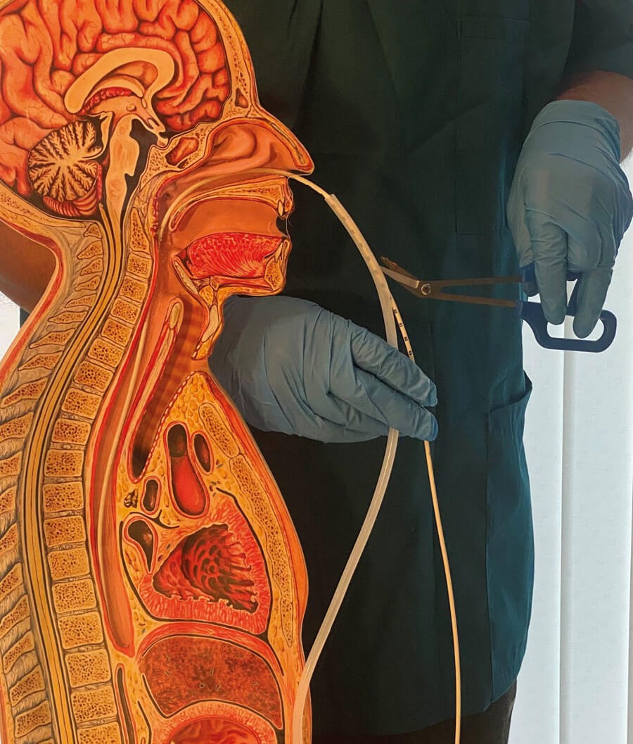
Figure 8a: The silicone conduit is split longitudinally using specialised scissors and removed while the new NG tube remains in place.
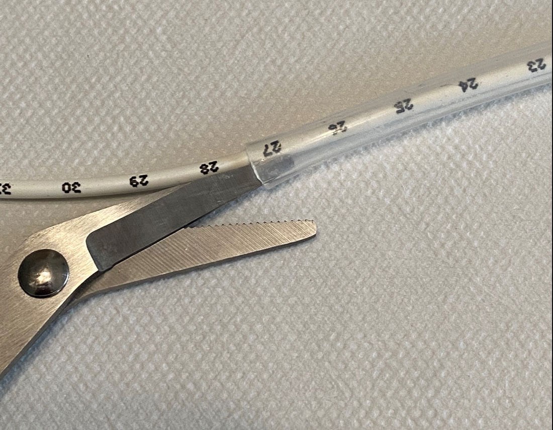
Figure 8b: Close-up view of scissors cutting the silicone conduit without damaging the NG or Ryle’s tube.
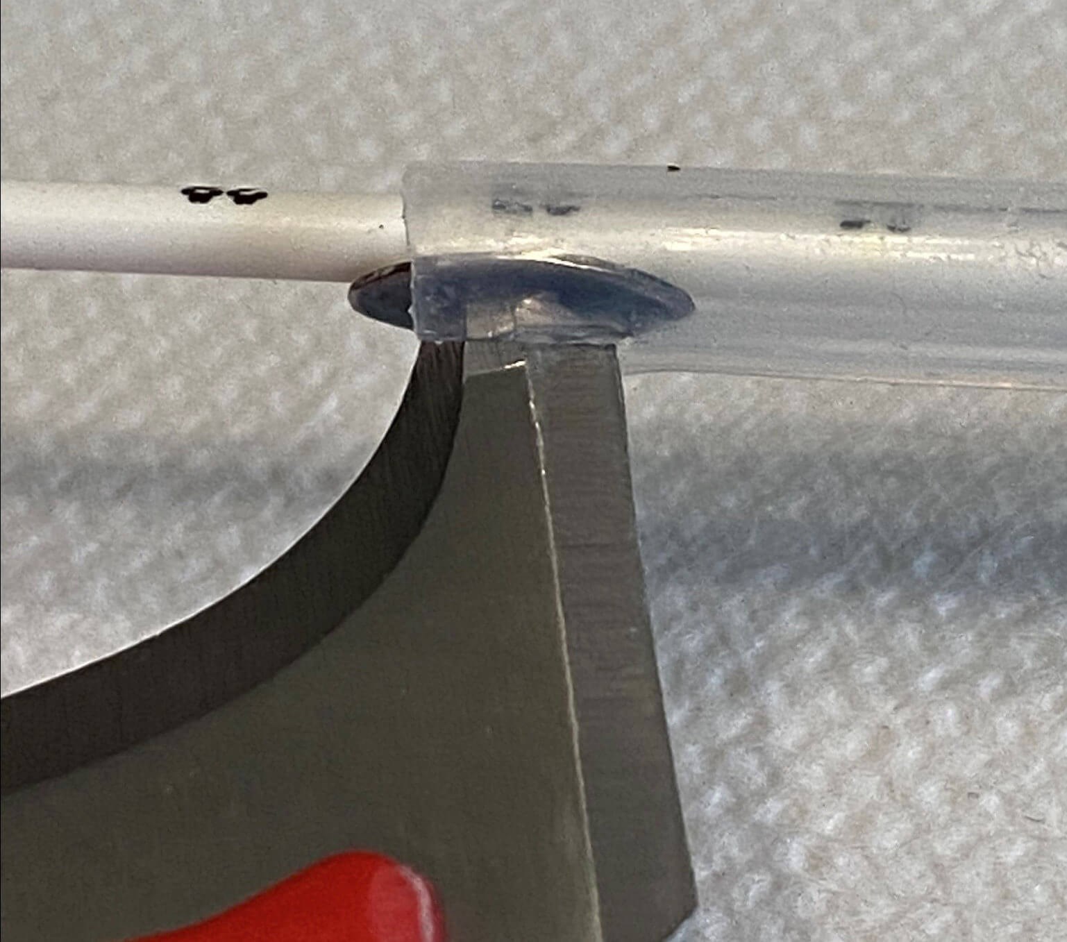
Figure 8c: A specialised blade is used to cut the silicone conduit without damaging the NG tube.
A medical-grade silicone conduit of appropriate calibre is lubricated and threaded over a thin, rigid, 60-cm-long forceps. The proximal connector of the in-situ NG or Ryle’s tube is trimmed to expose an accessible end, which is grasped and locked in the toothed jaws of the forceps. Using the in-situ tube as a leader, the silicone conduit is advanced gently over it until the distal end of the conduit lies at the gastric cardia. The conduit is held in position while the old NG or Ryle’s tube is withdrawn through it. With the conduit maintained from nostril to stomach, a new NG/Ryle’s tube is passed through its lumen and advanced to the required depth. The new tube is then stabilised while the conduit is withdrawn. During withdrawal the conduit is split longitudinally with dedicated scissors or a blade and separated, so that it can be removed without displacing or damaging the new tube. The new tube is left seated within the established tract.
As the conduit must slide over the in-situ tube and then accommodate the replacement tube, its calibre exceeds that of the tube being exchanged. This might have implications for tissue trauma in rare cases of very tight stricture, severe DNS or similar conditions that a rigid model cannot show (see Discussion). Although theoretically the new NG/Ryle’s tube should go back in the same position, the position needs to be confirmed by standard methods, including pH testing of aspirate and radiography, in line with national guidance.
Discussion
The Navigated Atraumatic Guidance (NAG) technique for reinsertion of an NG or Ryle’s tube uses the old failing tube itself as a guide, combined with a splittable conduit that holds the whole track open during the swap. Theoretically, this method appears to be a simple, safe, economical and reproducible method for reinsertion of an NG or Ryle’s tube. The technique does not remove the need for placement confirmation. It relocates a new tube along an assumed tract but it does not verify that the tract still ends in the stomach. pH testing and radiographic confirmation, interpreted by a competent operator, remain essential safeguards against the Never Event [2–4].
The same conduit-exchange principle might apply to other indwelling tubes where an established tract is worth preserving, but that too is speculative and would need its own evaluation. If feasibility is confirmed, the reasonable next steps are cadaveric assessment, then a supervised in-human feasibility series with defined success and complication endpoints and a comparator, before any claim of benefit.
Conclusion
The NAG technique is a repeatable method, on a rigid anatomical model, for exchanging an NG or Ryle’s tube along an existing tract. This report shows feasibility only. It does not establish safety, efficiency or benefit in patients, and standard confirmation of gastric placement is still required after any exchange. Cadaveric and then in-human evaluation with defined endpoints would be needed before clinical use.
References
1. Sparks DA, Chase DM, Coughlin LM, Perry E. Pulmonary complications of 9931 narrow-bore nasoenteric tubes during blind placement: a critical review. JPEN J Parenter Enteral Nutr 2011;35(5):625–9.
2. National Patient Safety Agency. Reducing the harm caused by misplaced nasogastric feeding tubes in adults, children and infants. Patient Safety Alert NPSA/2011/PSA002. London: NPSA; 2011.
3. NHS Improvement. Nasogastric tube misplacement: continuing risk of death and severe harm. Patient Safety Alert NHS/PSA/RE/2016/006. London: NHS Improvement; 2016.
4. Healthcare Safety Investigation Branch. Placement of nasogastric tubes. Investigation report. London: HSIB; 2020.
www.hssib.org.uk/patient-safety-investigations/
placement-of-nasogastric-tubes/investigation-report/
[Last accessed October 2026].
5. Vakil H, Lagzouli A, Lowe E. When the tube won’t go: strategies for difficult nasogastric intubation. Cureus 2025;17(11):e96725
6. Moharari RS, Fallah AH, Khajavi MR, et al. The GlideScope facilitates nasogastric tube insertion: a randomized clinical trial. Anesth Analg 2010;110(1):115–8.
7. Kirtania J, Ghose T, Garai D, Ray S. Esophageal guidewire-assisted nasogastric tube insertion in anesthetized and intubated patients: a prospective randomized controlled study. Anesth Analg 2012;114(2):343–8.
Disclosures Device status:
The 60-cm rigid forceps, the silicone conduit and the splitting blade are off-the-shelf items. Ethics: Bench/model work generally does not require ethical approval.
Declaration of competing interests: None declared.