Continuous intraoperative neural monitoring enhances thyroid surgery safety, helping predict and prevent recurrent laryngeal nerve injury in real time.
Recurrent laryngeal nerve (RLN) injury with resultant vocal cord paralysis (VCP) can have a significant impact on patient quality of life and remains the leading cause of medico-legal litigation following thyroid surgery. While direct visualisation of the nerve has long been the gold standard to prevent injury, the accuracy of predicting neural injury by visualisation alone is quite poor [1].
Intraoperative neural monitoring (IONM) has gained widespread use among thyroid surgeons for a variety of reasons, including: (A) intraoperative decision-making through prognostication of neural function, especially important in bilateral surgery; (B) early nerve identification and mapping; (C) dissection of the nerve; (D), understanding of surgical anatomic variation and branching; and (E) resident education [1].
I-IONM
Most literature involving IONM of the RLN is based on intermittent IONM data (I-IONM) and involves intermittent direct stimulation of the nerve by a handheld probe with a corresponding evoked laryngeal electromyographic (EMG) response. I-IONM is typically performed using endotracheal tube (ETT)-based electrodes at the level of the vocal cords. The use of IONM is widespread and continues to increase, rising from approximately 62.5% to 75.9% in the United States between 2016 and 2022 based on a recent retrospective cohort analysis of NSQIP thyroidectomy cases by Hearn et al [2]. However, the literature remains controversial regarding the ability of IONM to reduce RLN injury. While Hearn et al did show a small protective effect against unilateral RLN injury with the use of IONM in thyroidectomy (5.7% versus 6.5%), other studies have failed to show a clear advantage of IONM over visualisation alone [2]. Guidelines focused on I-IONM have been established by the International Neural Monitoring Study Group (INMSG) to improve the quality of IONM through standardisation of equipment setup and use [1]. However, a key limitation of I-IONM is the difficulty in predicting (and therefore preventing) an impending neuropraxic injury, such as a stretch or traction injury, which are the most common mechanisms of neural injury in thyroid surgery [3]. On the other hand, I-IONM has the potential to eliminate the risk of bilateral RLN injury with staging of bilateral thyroid surgery in response to a loss of EMG signal.
"Recurrent laryngeal nerve injury with resultant vocal cord paralysis can have a significant impact on patient quality of life and remains the leading cause of medico-legal litigation"
C-IONM
Continuous IONM (C-IONM) of the RLN in thyroidectomy was developed to overcome the inherent limitation of I-IONM in being able to detect a nerve injury occurring between intermittent neural stimulations. C-IONM provides real-time uninterrupted EMG data to allow for prediction of impending neuropraxic injury [4]. This allows the surgeon to perform corrective manoeuvres to prevent loss of EMG signal and resultant VCP.
Both C-IONM and I-IONM require the same multichannel EMG system, ETT surface electrodes, neural stimulator probe and display; C-IONM adds a continuous vagal stimulation electrode and the corresponding ground electrode [4]. Once the patient is intubated with an ETT with laryngeal EMG electrodes (following the 2011 INMSG Guidelines), thyroidectomy proceeds with initial exposure of the carotid sheath on the ipsilateral side of surgery. Direct stimulation of the vagus nerve (VN) with the handheld probe ensures an intact pre-dissection VN signal, confirming appropriate equipment setup and functionally intact RLN along its entire length. At this point, gentle 360-degree dissection over a short segment of the VN is then performed for application of the C-IONM electrode. While different types of electrodes are available, examples of equipment setup and electrode placement are shown in Figures 1 and 2.
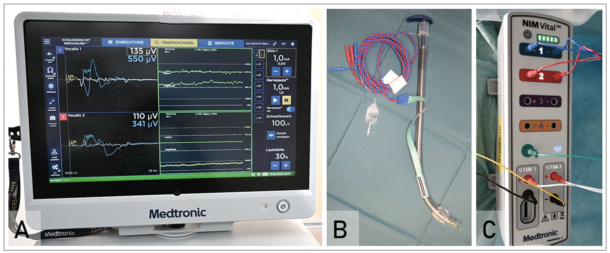
Figure 1: View of components of intraoperative neuromonitoring.
A. NIM Vital™ Nerve Monitor screen during continuous neuromonitoring.
B. NIM EMG tube with recording endotracheal tube surface electrodes.
C. Connecting NIM Vital™ interface box between recording tube surface electrodes and NIM Vital™ Nerve Monitor.
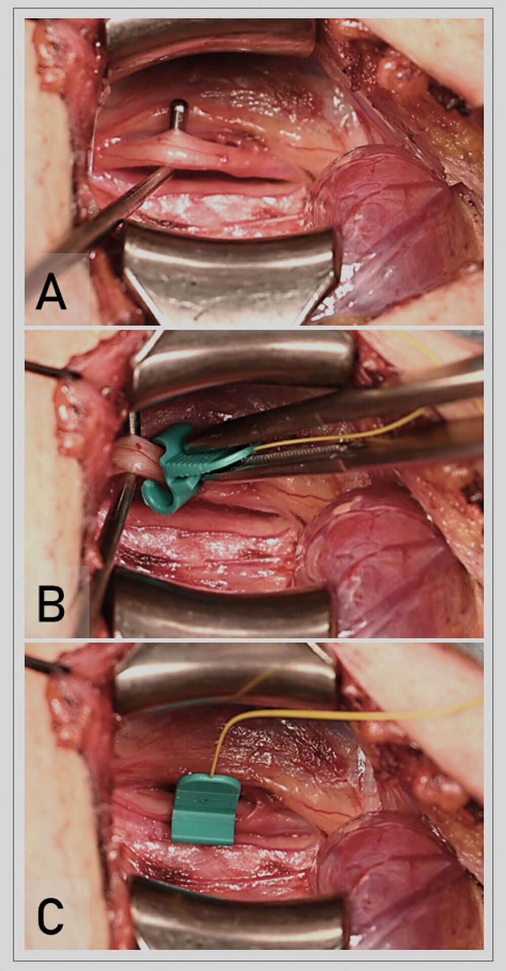
Figure 2: Placement of the monopolar APS® electrode onto the vagus nerve before thyroid surgery.
A. View of the left carotid sheath depicting the common carotid artery and internal jugular vein, with the vagus nerve held in position by a nerve retractor.
B. Positioning of the APS® electrode with a forceps on the dissected segment of the vagus nerve from a 45° angle.
C. View of the APS® electrode resting on the vagus nerve inside the carotid sheath.
Typical initial settings for C-IONM include a current of 1 mA and frequency of 1 Hz (with vagal stimulation occurring every one second). While the frequency of stimulation may be adjusted based on surgeon preference, system calibrations should optimise maximum achievable baseline vagal amplitude, with a minimum amplitude of 500 µV deemed appropriate for a reliable EMG signal and interpretation of data. Complementary application of the direct handheld stimulation probe should still be employed for RLN identification and mapping, as during I-IONM.
"I-IONM has the potential to eliminate the risk of bilateral RLN injury with staging of bilateral thyroid surgery in response to a loss of EMG signal"
Once the baseline vagal EMG reference curve is established, changes in vagal amplitudes and latencies are displayed on a timeline on the EMG monitor. Audible and visual warning thresholds notify the surgeon when a manoeuvre may be causing nerve irritability or impending injury. Changes in EMG signal are categorised according to (A) non-dangerous changes, (B) dangerous changes (the ‘combined event’), and (C) loss of EMG signal (LOS, <100 µV) [4]. Non-dangerous EMG changes may occur with mechanical manipulation of the thyroid and frequently resolve immediately upon returning the thyroid to its original position. Isolated EMG amplitude changes without a latency shift may signal movement of the endotracheal tube and corresponding laryngeal electrodes. However, more worrisome EMG changes occur in the setting of a continued decrease in EMG amplitude combined with an increase in latency.
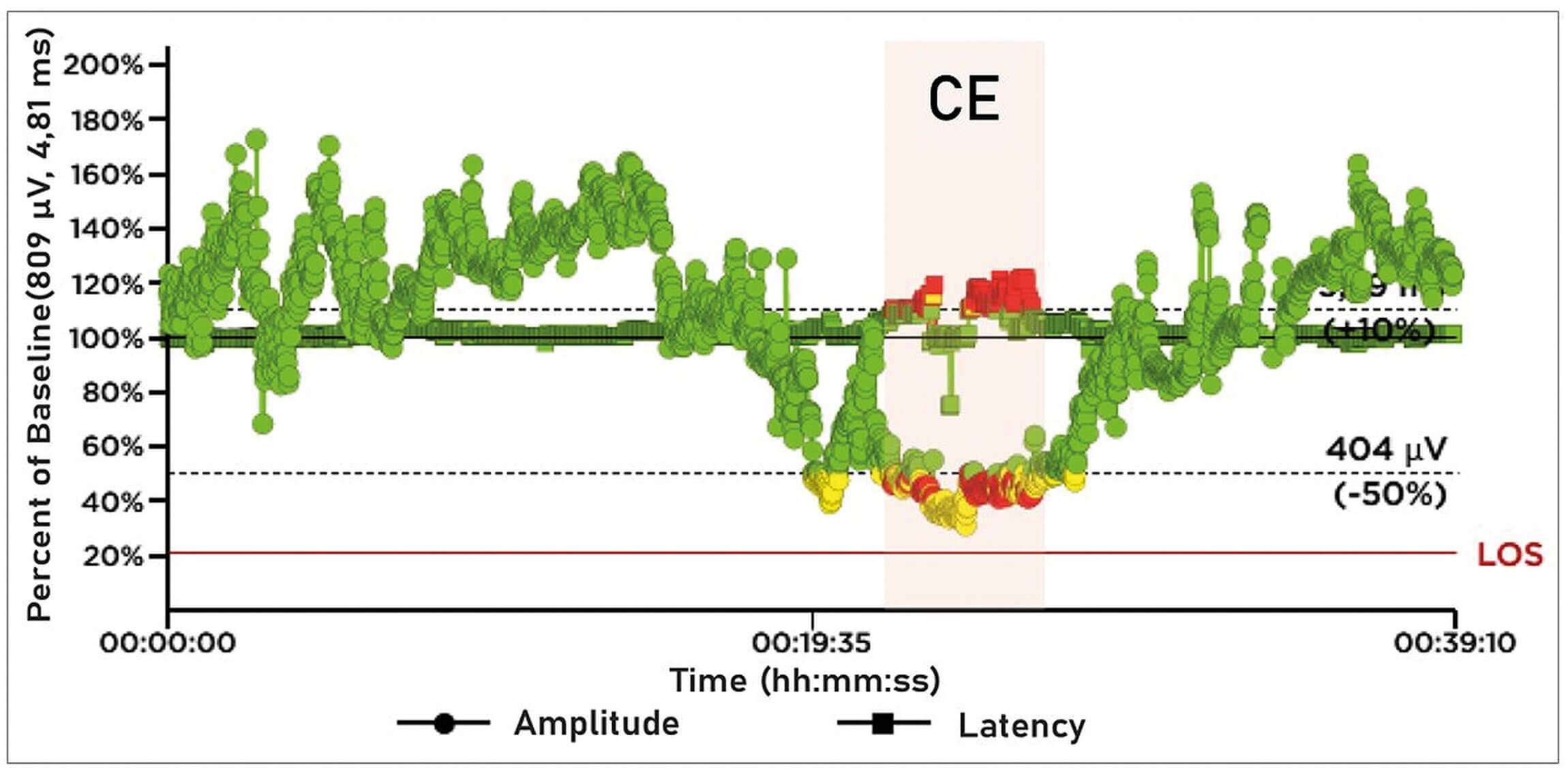
Figure 3: Electromyography (EMG) tracings showing a traction-induced combined event with amplitude <50% (but >100µV) and latency >110 % of baseline, signifying impending nerve injury in continuous neural monitoring. This is completely reversible after immediately resolving the causative manoeuvre.
An adverse ‘combined event’ (CE) is defined as an EMG amplitude decrease by >50% with a concordant increase in latency of >10% (Figure 3) [4]. Such adverse CEs have been associated with development of VCP, with a PPV of 33% and NPV of 97%. With immediate surgical correction, 73% were reversible and 80% did not result in postoperative VCP [4]. Incomplete recovery after loss of EMG signal is defined as amplitude increase to >100 microvolts but less than 50% of the baseline vagal amplitude, and is associated with an increased risk of postoperative VCP. Complete recovery after loss of EMG signal is defined as amplitude increase to >50% of the baseline. If intraoperative recovery has not occurred or is incomplete by 20 minutes, the risk of immediate postoperative VCP is high (94.6% of segmental [focal] RLN injuries and 69.5% of global injuries [where a focal point of injury cannot be identified]). This has significant implications for bilateral surgery, where a staged procedure is recommended when complete recovery of EMG amplitude is not seen by 20 minutes.
There is a learning curve to set up and interpret C-IONM data. One limitation of C-IONM includes intermittent dislocation of the vagal electrode during surgery, which does improve with surgeon experience. C-IONM is also more limited in predicting a segmental (focal) nerve injury, such as from cautery or transection, as these injuries tend to occur suddenly and irreversibly. In addition, rare incidences of hemodynamic instability or reversible vagal neuropraxia have been reported during C-IONM [5]. However, the majority of high-volume studies have shown safe use of C-IONM when stimulation currents and frequencies are kept low, including in paediatric and high-risk populations [4,5]. Low stimulation current of 1 to 2 mA do not activate the demyelinated C-fibres in the VN, which are responsible for the majority of autonomic effects [4].
Several studies suggest superiority of C-IONM over I-IONM in preventing RLN injury and resultant VCP after thyroidectomy. A comparative study of over 10,000 nerves at a tertiary care centre in Germany showed that use of C-IONM translated into a 1.7-fold decrease in early postoperative VCP rates and 30-fold decrease in permanent VCP rates compared to I-IONM [6]. A recent meta-analysis showed an overall 2.5% rate of VCP and permanent rate of 0.05%, which are lower than published data on I-IONM and visualisation alone [5].
"With immediate surgical correction, 73% were reversible and 80% did not result in postoperative"
Future directions
To bridge the gap between the more widely used I-IONM and the potentially superior C-IONM in terms of RLN injury in thyroidectomy, new technology is being studied that allows for tracking of neural function even with I-IONM (NIM NerveTrendTM, Medtronic, Jacksonville, Fl). Two single-centre randomised controlled trials compare NIM NerveTrendTM to either traditional I-IONM or C-IONM in terms of RLN injury rates and decreased need for staged surgery [7,8]. These studies suggest inferiority of traditional I-IONM compared to NIM NerveTrendTM and noninferiority of NIM NerveTrendTM compared to C-IONM.
Future work should focus on broader validation, cost-effectiveness and integration with machine-learning–based artifact reduction to reliably predict early postoperative nerve function. I-IONM and C-IONM monitoring should be used in a complementary manner in modern thyroid surgery whenever possible to optimally utilise the strengths of both [9].
References
1. Randolph GW, Dralle H, International Intraoperative Monitoring Study G, et al. Electrophysiologic recurrent laryngeal nerve monitoring during thyroid and parathyroid surgery: international standards guideline statement. Laryngoscope 2011;121 Suppl 1:S1–16.
2. Hearn M, You B, Mady LJ, et al. Progress and Outcomes of Intraoperative Nerve Monitoring During Thyroidectomy. JAMA Otolaryngol Head Neck Surg 2025;151(3):236–42.
3. Liddy W, Wu CW, Dionigi G, et al. Varied Recurrent Laryngeal Nerve Course Is Associated with Increased Risk of Nerve Dysfunction During Thyroidectomy: Results of the Surgical Anatomy of the Recurrent Laryngeal Nerve in Thyroid Surgery Study, an International Multicenter Prospective Anatomic and Electrophysiologic Study of 1000 Monitored Nerves at Risk from the International Neural Monitoring Study Group. Thyroid 2021;31(11):1730–40.
4. Schneider R, Randolph GW, Barczynski M, et al. Continuous intraoperative neural monitoring of the recurrent nerves in thyroid surgery: a quantum leap in technology. Gland Surg 2016;5(6):607–16.
5. Ku D, Hui M, Cheung P, et al. Meta-analysis on continuous nerve monitoring in thyroidectomies. Head Neck 2021;43(12):3966–78.
6. Schneider R, Machens A, Sekulla C, et al. Superiority of continuous over intermittent intraoperative nerve monitoring in preventing vocal cord palsy. Br J Surg 2021;108(5):566–73.
7. Barczynski M, Dworak M, Krakowska K, et al. Clinical Validation of NerveTrend vs. NerveAssure Mode of Intraoperative Neuromonitoring in Prevention of Recurrent Laryngeal Nerve Injury During Thyroid Surgery: A Randomized Controlled Trial. Ann Surg 2025;282(5):709–16.
8. Barczynski M, Konturek A. Clinical validation of NerveTrend versus conventional i-IONM mode of NIM Vital in prevention of recurrent laryngeal nerve events during bilateral thyroid surgery: A randomized controlled trial. Head Neck 2024;46(3):492–502.
9. Sinclair CF, Buczek E, Cottril E, et al. Clarifying optimal outcome measures in intermittent and continuous laryngeal neuromonitoring. Head Neck 2022;44(2):460–71.
Declaration of competing interests: GWR received research grants from Eisai, Medtronic and Getinge, and received consulting fees from Medtronic and Eisai.