
Stricture after total laryngectomy is common but not inevitable. New insights into risk factors could help clinicians anticipate problems and improve swallowing outcomes.
Why this complication deserves renewed attention
Total laryngectomy remains one of the defining operations in head and neck oncology. Although the procedure can offer durable oncological control, the modern question is no longer survival alone, but what quality of life after treatment looks like. In that context, pharyngo-oesophageal stricture deserves more attention. It is not a rare technical inconvenience; it is a clinically meaningful source of dysphagia, repeated intervention, nutritional compromise and reduced quality of life [1,2].
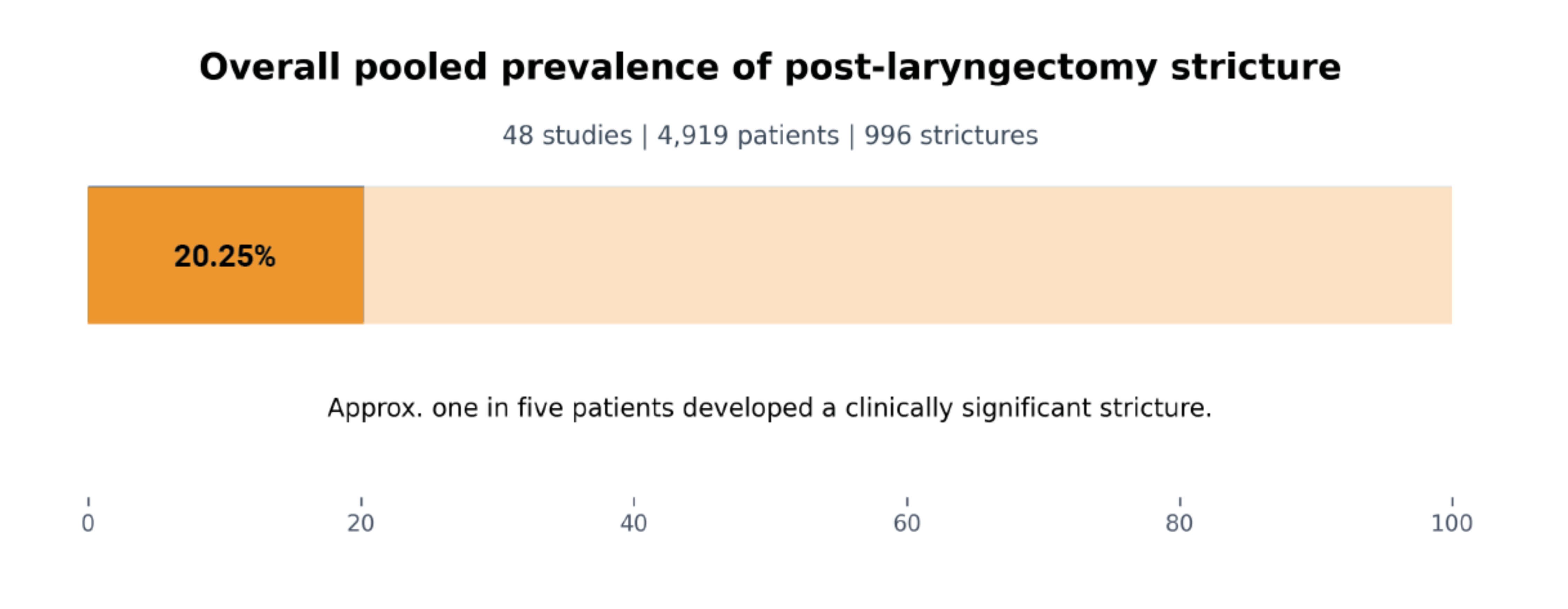
Figure 1: Overall pooled prevalance of post-laryngectomy stricture. Across 48 studies and 4919 patients.
Our recent systematic review and meta-analysis found that approximately one in five patients developed a clinically significant stricture following total laryngectomy. As shown in Figure 1, the overall pooled prevalence was 20.25%. Across 48 studies and 4,919 patients, the pooled stricture rate was 20.25%, which represents 996 strictures [1]. This is a sobering figure. For a complication that can require serial dilatations, prolong swallowing dysfunction and shape the patient’s experience of recovery, that burden is substantial. It also means that stricture formation should be thought of as a central functional outcome of laryngectomy care rather than a secondary postoperative issue [1,2].
Risk is not random
The principal contribution of the review is that it moves the discussion away from isolated anecdotes and toward a recognisable pattern of risk. Post-laryngectomy stricture is multifactorial. It reflects treatment history, tumour anatomy, reconstructive burden, surgical technique and, to some extent, patient-level healing factors. The main messages are summarised in Table 1 [1].
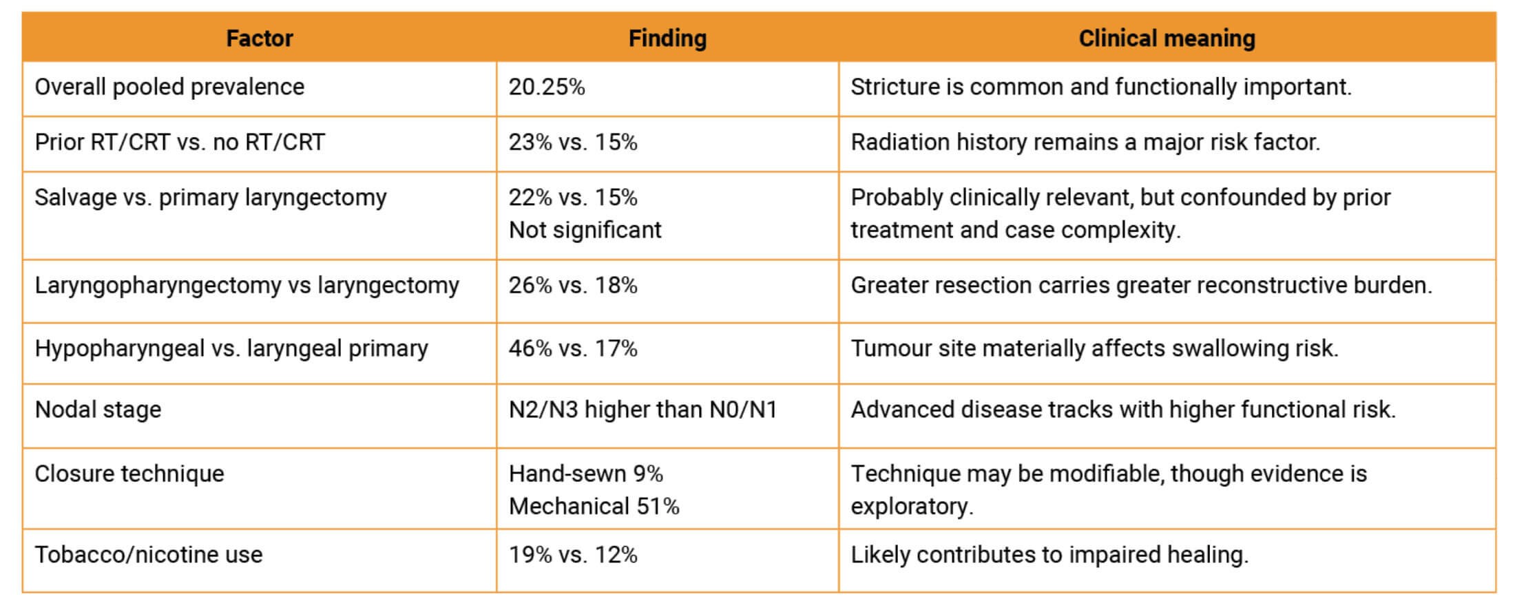
Table 1. Main clinical takeaways from the systematic review.
Prior radiation or chemoradiation emerged as one of the clearest associations. Patients with a history of radiotherapy (RT) or chemoradiotherapy (CRT) had higher stricture rates than those without prior radiation exposure, at 23% versus 15% [1]. That finding is biologically plausible. Radiation impairs microvascularity, tissue elasticity and mucosal healing, and the broader reconstructive literature has long described these effects as drivers of compromised wound repair [3]. Importantly, this helps explain why salvage settings remain clinically high-risk even when salvage timing itself does not always achieve statistical significance in pooled comparisons [1]. Rather than viewing salvage laryngectomy as a single category, it is more useful to recognise how prior radiation and the extent of resection interact in the same patient [1,3].
"Stricture formation after total laryngectomy is common, clinically important and patterned enough to inform better decision-making"
Anatomy and extent of resection matter
The second major message is that anatomy matters. Patients undergoing laryngopharyngectomy had higher stricture rates than those undergoing laryngectomy alone, at 26% versus 18% [1]. Tumour site carried an equally strong signal. Hypopharyngeal primaries were associated with substantially higher stricture rates than laryngeal primaries, at 46% versus 17% when controlling for RT/CRT [1]. Advanced nodal disease also linked to worse outcomes, with both N2 and N3 disease associated with significantly higher stricture prevalence than N0 or N1 disease [1].
These findings make intuitive clinical sense. Greater pharyngeal resection means less native mucosa, more complex reconstruction and a greater risk of suture-line tension. Hypopharyngeal disease, in particular, is not simply a different anatomical label; it often represents a different reconstructive problem altogether. Earlier clinical series in non-radiated laryngeal and hypopharyngeal carcinoma also pointed toward the importance of anatomical burden and postoperative complications in shaping recovery, and comparative work has shown that function is generally less favourable when reconstruction extends into the pharynx [4,5]. For clinicians, this reframes stricture not as an unpredictable late event, but as a foreseeable consequence of specific anatomical and oncological factors. Figure 2 therefore emphasises the gradient in risk across prior RT/CRT, extent of resection, tumour site and selected patient-level factors [1].
"If closure method and reconstructive choice influence downstream swallowing outcomes, then stricture prevention begins in the operating theatre"
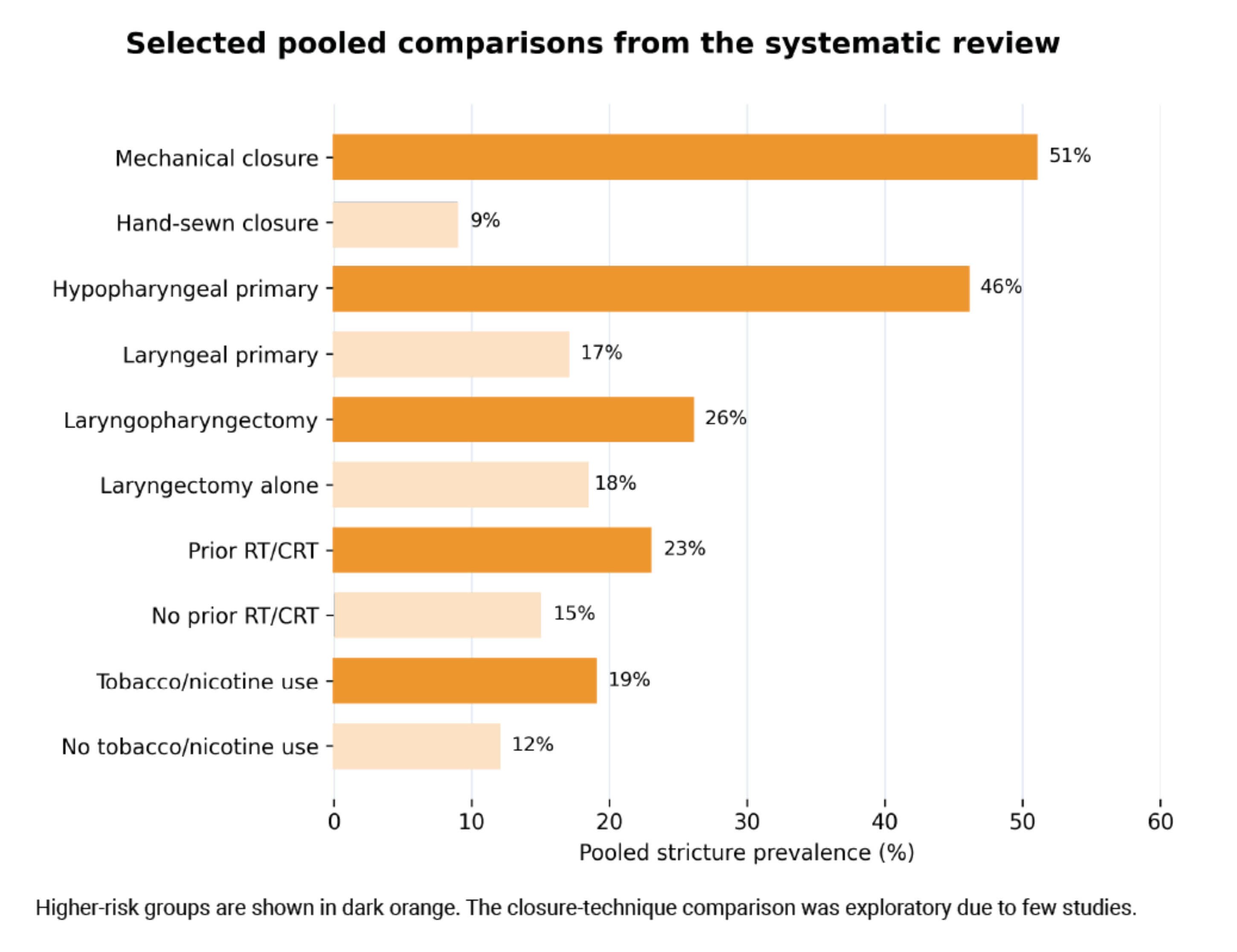
Figure 2: Selected pooled comparisons from the systematic review. Higher-risk groups are shown in dark orange.
Technique may be one of the few modifiable levers
Perhaps the most practice-shaping observations in the review relate to operative technique. Hand-sewn closure was associated with substantially lower stricture rates than stapled (mechanical) closure, at 9% versus 51%, although the authors appropriately considered this analysis exploratory because relatively few studies reported closure technique in a way suitable for pooling [1]. Similarly, reconstruction type mattered in selected comparisons: fasciocutaneous free flaps had the highest stricture prevalence, while visceral free flaps had the lowest, with a significant difference when those two categories were compared directly [1].
"Stricture is not simply a late complication to manage; it is a risk to anticipate"
These findings must be interpreted cautiously because confounding by indication is difficult to exclude. Stapled closure may be used in different clinical circumstances, and flap choice is influenced by patient and reconstructive factors. Even so, the signal is clinically provocative. If closure method and reconstructive choice influence downstream swallowing outcomes, then stricture prevention begins in the operating theatre. The review also found a modest association between tobacco or nicotine use and stricture risk, with rates of 19% versus 12% in non-users, although this became borderline after adjustment for RT/CRT [1]. That pattern is consistent with a wider wound-healing literature in which tissue perfusion, collagen remodelling and local inflammation influence scar formation and luminal narrowing [3]. Alcohol use and voice restoration timing, by contrast, did not show significant pooled effects in the available data [1].
What should clinicians do with these findings?
The immediate clinical value of the review lies in risk stratification. Patients with prior RT/CRT, hypopharyngeal disease, advanced nodal stage and more extensive resections should be counselled that their functional risk is higher [1]. That does not mean poor swallowing is inevitable, but it does mean that postoperative expectations, surveillance and rehabilitation should be adjusted accordingly. For the multidisciplinary team, this may justify earlier speech and language therapy input, closer nutritional follow-up and a lower threshold for instrumental assessment when swallowing begins to deteriorate [1,2].
One gap in the literature is the absence of agreed surveillance pathways for stricture in the laryngectomy population. At present, many intervene reactively once dysphagia is established. A more structured approach to follow-up, particularly in higher-risk subgroups, may prove beneficial, although prospective data are still needed [1,2].
From association to prevention
This review does not prove causation and it does not eliminate the heterogeneity that characterises laryngectomy outcomes research. Many included studies were retrospective, definitions of stricture varied and some subgroup analyses were exploratory [1]. Even so, the direction of travel is clear. Stricture formation after total laryngectomy is common, clinically important and patterned enough to inform better decision-making.
The larger lesson is that functional outcome should be designed for, not merely measured after the fact. If we know that prior radiation, greater reconstructive burden and specific technical choices are linked to later dysphagia, then prevention should become part of routine operative thinking. That is the most important takeaway from the review. Stricture is not simply a late complication to manage; it is a risk to anticipate [1].
References
1. Stellern J, Corbisiero MF, Burnet G, et al. Risk Factors for Esophageal Stricturing Status Post Total Laryngectomy: A Systematic Review and Meta-analysis. Dysphagia 2026 [ePub ahead of print].
2. Petersen JF, Pezier TF, van Dieren JM, et al. Dilation after laryngectomy: incidence, risk factors and complications. Oral Oncology 2019;91:107–12.
3. Jacobson LK, Johnson MB, Dedhia RD, et al. Impaired wound healing after radiation therapy: a systematic review of pathogenesis and treatment. JPRAS Open 2017;13:92–105.
4. Herranz J, Sarandeses A, Fernandez MF, et al. Complications after total laryngectomy in nonradiated laryngeal and hypopharyngeal carcinomas. Otolaryngol Head Neck Surg 2000;122(6):892-8.
5. Gadepalli C, De Casso C, Silva S, et al. Functional results of pharyngo-laryngectomy and total laryngectomy: a comparison. J Laryngol Otol 2012;126(1):52–7.
Declaration of competing interests: None declared.