Plastic temporal bones transformed ear surgery training, letting trainees practise complex procedures safely before operating on patients.
I attended the Stell and Maran’s surgical course in the early 70s. It was superb but, with the confidence of naivety, I thought I could do better. If I ever ran a course, simulation surgery would complement the theoretical lectures. It seemed to me that practical surgery was never formally taught, never examined, and often neglected.
It was thought that if one was good at passing theoretical exams, practical surgical skill could be taken for granted. Having watched some senior surgeons at work, I knew this presumption did not hold true. How can it be that practical skill, so vital to success – as in the case of a concert pianist – is repeatedly examined, yet surgeons never take an exam that demonstrates their manual dexterity in surgery?
I started the Glasgow Temporal Bone Course in 1976. I think this was the first practical course in ear surgery in the UK and maybe the first practical course in all UK surgery. Initially we used cadaver bones which were a wonderful simulator for introducing aspiring ENT surgeons to the challenges of otological surgery. However, cadaver bones were so good that I had the uncomfortable feeling that at some time in the future they would become unavailable. They were too good to be true.

Alastair in his workshop.
Sometime during the 80s I was at neighbour’s barbecue, chatting to the host who was a senior lecturer in dentistry. He was telling me how he had spent the day teaching dental students to drill teeth on a plastic model of the mouth. I scoffed that ear anatomy was so complicated and had so many tiny and vital structures that we had to use cadaver bones. In the ensuing weeks, I regretted my offhand retort to my host. Would it indeed be possible to make a plastic temporal bone?
"It seemed to me that practical surgery was never formally taught, never examined, and often neglected"
I had a dried dissected part of a temporal bone showing the opened semicircular canals, and wondered, how can I make a copy of this? My first effort involved a paste used in filling dents in car bodies. That didn’t work. I tried various other materials and still made no progress. By chance, I attended a craft fair in the local town hall. There were wood carvings of flying ducks, blown glass decorations, etc., but one item caught my attention: a plaster cast of a foot. Apparently, the douce folk of Edinburgh decorate their mantlepieces with them.
As luck would have it I had the dissected bone in my pocket and I asked owner of the stall if he could make a cast of it. He had a good look and said he would try. Ten days later I received a beautiful cast, not in plaster but in polyurethane, with the canals finely defined. Yippee! This man had the skills I had long been looking for. I thought I would test him again and sent him a set of ossicles. Back came a perfectly cast set. The stapes was a special delight, I thought it would be too small and delicate to cast.
I now knew someone who could make casts of parts of temporal bone, but how could I make a complete bone? I realised I did not need to make casts of the cochlear or labyrinthine ducts, since skill in these areas is required only of very senior surgeons, if at all. It was clear to me that I would have to make two large casts of the temporal bone: one that represented the cortex and mastoid air cells, and one that represented the medial wall of the tympanum and excavated mastoid. The marrying of these two components would be disguised in the tympanomastoid and tympanosquamous sutures of the external auditory canal. Each ossicle would be cast separately and skin of the EAC and the tympanic membrane would be made in latex from solid models of the EAC. The horizontal part of the facial canal was uncapped and the vertical part had been opened during the mastoid dissection. Thus, the whole course of the facial nerve through the ear could be easily and precisely filled with silicone rubber.

A set of ossicles.
One problem which caused great difficulty was the pneumatisation of the mastoid. I spent months seeking a solution to this feature. I even tried effervescent liver salts to see if its bubbles would do the trick. I then had inspiration. I made a cast where the pneumatised area was a solid block and then I drilled holes in it and made a mould from this drilled cast. From this mould, I could make daughter casts with a pneumatised mastoid – problem solved!
In the meantime, the Glasgow Temporal Bone Course continued to be successful, drawing 40 participants each year. Practical dissection and theoretical tutorials over three days seemed to be attractive to the aspiring ear surgeons. In the middle 90s it became clear we could no longer obtain cadaver bones. Fortunately at that time I thought my plastic bones had become ‘good enough’. Plastic bones were happily accepted by the next 400 participants on the course and by my colleagues, Iain Swan and George Browning.

Atticotomy.

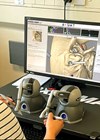
Bone 2 in operating position.
In the middle 00s, I retired from clinical practice but continued to make and sell what were now Pettigrew Temporal Bones. Business was encouraging and enormously satisfying. I got a thrill out of the idea that people worldwide would pay for something I had conjured up in my head and made with my hands. But technology does not stand still. Computers were becoming much more clever and provided magnificent images that could be worked on with haptic devices. Printed 3D temporal bones were being made and sold. However, the new technologies had their limitations [1]. Moreover, I kept making minor improvements in the design and manufacture of my bones. It seemed to me that my bones allowed the aspiring surgeon to do the complete range of operations needed in the first five years of independent practice – namely myringotomy, ventilation tube insertion, raise tympanomeatal flap, myringoplasty, ossiculoplasty, stapedotomy and prosthesis insertion, posterior tympanotomy, canal wall up/down mastoidectomy and facial nerve exposure. In addition, pathology such as perforation of the tympanic membrane, fixity of the stapes footplate and cholesteatoma can be incorporated.
"Ten days later I received a beautiful cast, not in plaster but in polyurethane, with the canals finely defined. Yippee!"
The bones are designed to equip the aspiring surgeon with a mental 3D image of the temporal bone so they are able to predict what vital structure will be found just beyond the point of their instrument. For a beginner, fully dissecting a Pettigrew bone requires about five hours. I estimate that about 40 hours of practice on temporal bones are required before an average aspiring surgeon can begin to operate on a patient under the direct supervision of an expert.

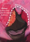
Perforation of tympanic membrane.
Cadaver temporal bones are the gold standard simulator for trainee ear surgeons but, in contradistinction to Pettigrew Temporal Bones, there are legal limitations to use and supply is limited. It is wonderful that nowadays trainee ear surgeons have four or more devices to choose from and that each one will have its place in providing the trainee with practical skills that will make them a safe, confident and happy surgeon.
Reference
1. Frithioff A, Frendø M, Weiss K, et al. Effect of 3D-Printed Models on Cadaveric Dissection in Temporal Bone Training. OTO Open 2021;5(4):2473974X2110650.
Declaration of competing interests: None declared.