ENT features archive for May 2014
Thyroid nodules – time for a rational imaging approach
“The more you know, the harder it is to take decisive action. Once you become informed, you start seeing complexities and shades of gray. You realize that nothing is as clear as it first appears. Ultimately, knowledge is paralyzing.” Calvin,...
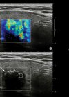
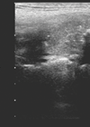
Thyroid ultrasound elastography: does nodule stiffness predict malignancy?
Approximately 50% of the general population has a thyroid nodule while 5-15% of these are malignant [1]. A major challenge, therefore, is how to detect the malignant nodules for appropriate, timely treatment and avoid unnecessary, costly investigations for the remainder....
Radiology and sinus disease: “the ever-evolving landscape”
Computed tomography (CT) remains the imaging modality of choice in assessment of patients with symptoms of chronic rhinosinusitis resistant to conservative treatment. In the last 10 years, CT technology has seen significant advances with the development and integration of multi-detector...
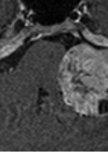
Imaging tinnitus
Tinnitus is a common sensation with a reported prevalence of 7-32%. The British National Study of Hearing recorded that 10% of adults suffered from prolong spontaneous tinnitus, and approximately a quarter of these are subsequently referred to hospital for investigation...
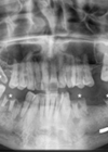
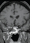
Radiology of referred otalgia
Otalgia is a common presenting complaint to Ear Nose and Throat Departments. Otalgia is either primary or secondary (referred) [1]. Referred otalgia is a ‘red flag’ symptom and can be a diagnostic challenge for clinicians and radiologists as the pathology...
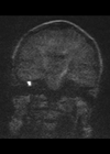
Detecting postoperative cholesteatoma with diffusion weighted magnetic resonance imaging
Middle ear cleft cholesteatoma is an inflammatory disease that erodes local bony structures and can cause otorrhea, hearing loss, vertigo and intracranial complications. It is usually treated with surgery, typically canal wall up (CWU) or canal wall down (CWD) surgery....
Imaging in hyperparathyroidism
Following their caudal migration at eight weeks of development, the parathyroid glands normally locate posterolaterally to the upper pole of the thyroid gland at the level of the cricoid cartilage (superior parathyroid glands arising from the fourth branchial pouch and...
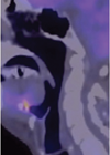
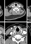
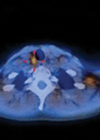
Head and neck cancer and PET-CT
Positron emission tomography-computed tomography (PET-CT) is an imaging technique in which abnormalities of tissue metabolism are precisely superimposed onto the anatomy. It relies on the premise that malignant cells are more metabolically active compared with non-malignant cells. On this basis,...
Nuclear heads – and necks
Imaging of the extra-cranial head and neck is challenging due to the anatomic complexity of the region. CT, MRI and ultrasonography (US) are amongst the most frequently utilised radiological modalities in head and neck imaging but do not always provide...
Imaging and management of head and neck vascular anomalies
Vascular anomalies are a diffuse spectrum of abnormalities which often involve the head, neck and oral cavity. They are frequently misnamed, often being generically labelled as haemangiomas. This lack of basic understanding can cause confusion leading to a cascade of...
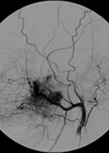
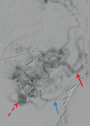
Role of interventional neuroradiology in otorhinolarygological pathology – a brief review
Introduction Since its advent in 1964 when Dotter percutaneously dilated a stenosed femoral artery [1], interventional radiology has undergone tremendous advancement in both imaging and devices that have enabled the operator (interventional radiologist) to access very distal small vasculature and...